Japanese
English
- 有料閲覧
- Abstract 文献概要
- 1ページ目 Look Inside
われわれは胃角上部前壁に存在した,粘膜内にとどまる陥凹型早期胃癌で,興味ある組織形態を示し,術前の深達度診断が困難であった症例を経験した.本症例は胃底腺粘膜領域に発生した低分化型癌で,悪性サイクルを示したので,これらについて若干の文献的考察を加え報告する.
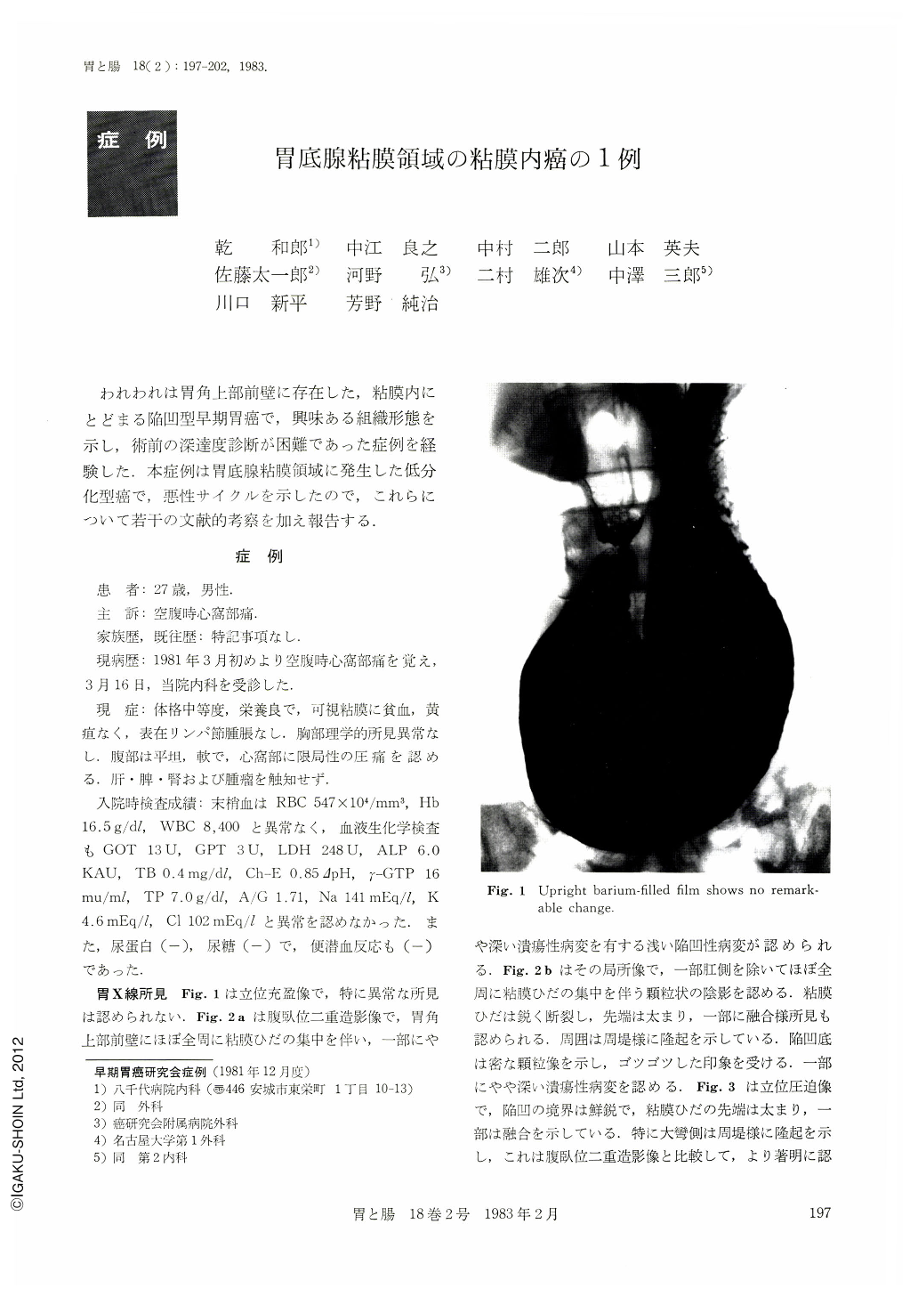
A 27-year-old man visited our hospital with complaint of hunger epigastralgia. X-ray examination showed clearly the shallow depressed area with converging folds on the anterior wall of the lower gastric body, in prone double contrast film and upright compression film. The mucosal convergence revealed abrupt interruption and the tips of the mucosal folds were clubbed and seemed to be fused. The granular mucosa was seen in the depressed area and the small ulcer in it. The initial endoscopy revealed the small ulcer was covered with a white coat. The second endoscopy showed the small ulcer had changed into a scar. The lesion was diagnosed as advanced cancer like Ⅱc+Ⅲs type. The biopsy specimens confirmed signet-ring cell carcinoma. In the resected specimen, the lesion, 7×14 mm in diameter, was located in the fundic gland area. Histologically, poorly differentiated adenocarcinoma and signet-ring cell carcinoma were found in the submucosal layer.
This case seemed to be at one phase of the so-called malignant cycle. The cancerous invasion was found in the mucosal layer within the limits of the edge of the depressed area covered with regenerative epithelium. By x-ray examination, the granular lesion in the Ⅱc area was visualized as a deep cancerous invasion, but retrospectively, the size of the granules seemed to be almost equal. The cancer in the fundic gland area has a tendency of making severe fibrosis in the submucosal layer. The fibrosis of this case was not so severe but it maked us miss the diagnosis. This case made evident the difficulty of the diagnosis of the extent of cancer in the fundic gland area.

Copyright © 1983, Igaku-Shoin Ltd. All rights reserved.


