Japanese
English
- 有料閲覧
PPV購入案内
1,320円 (1,200円+税10%) こちらは、255ページから260ページの文献です。購入には医書.jp本会員登録が必要です。
会員登録完了後に改めて上記「購入サイトへ」を選択してください。
または「購入サイトへ」選択後に「購入ログイン」、「新規会員登録」の順に進んでください。
会員登録について詳しくはをご確認ください。
- Abstract 文献概要
- 1ページ目 Look Inside
胃底腺粘膜から発生したⅡc+Ⅲ型の粘膜内癌で,術前深達度診断がsmまたはsm以深に浸潤していると診断された症例を経験し,その深達度診断について検討を加えたので報告する.
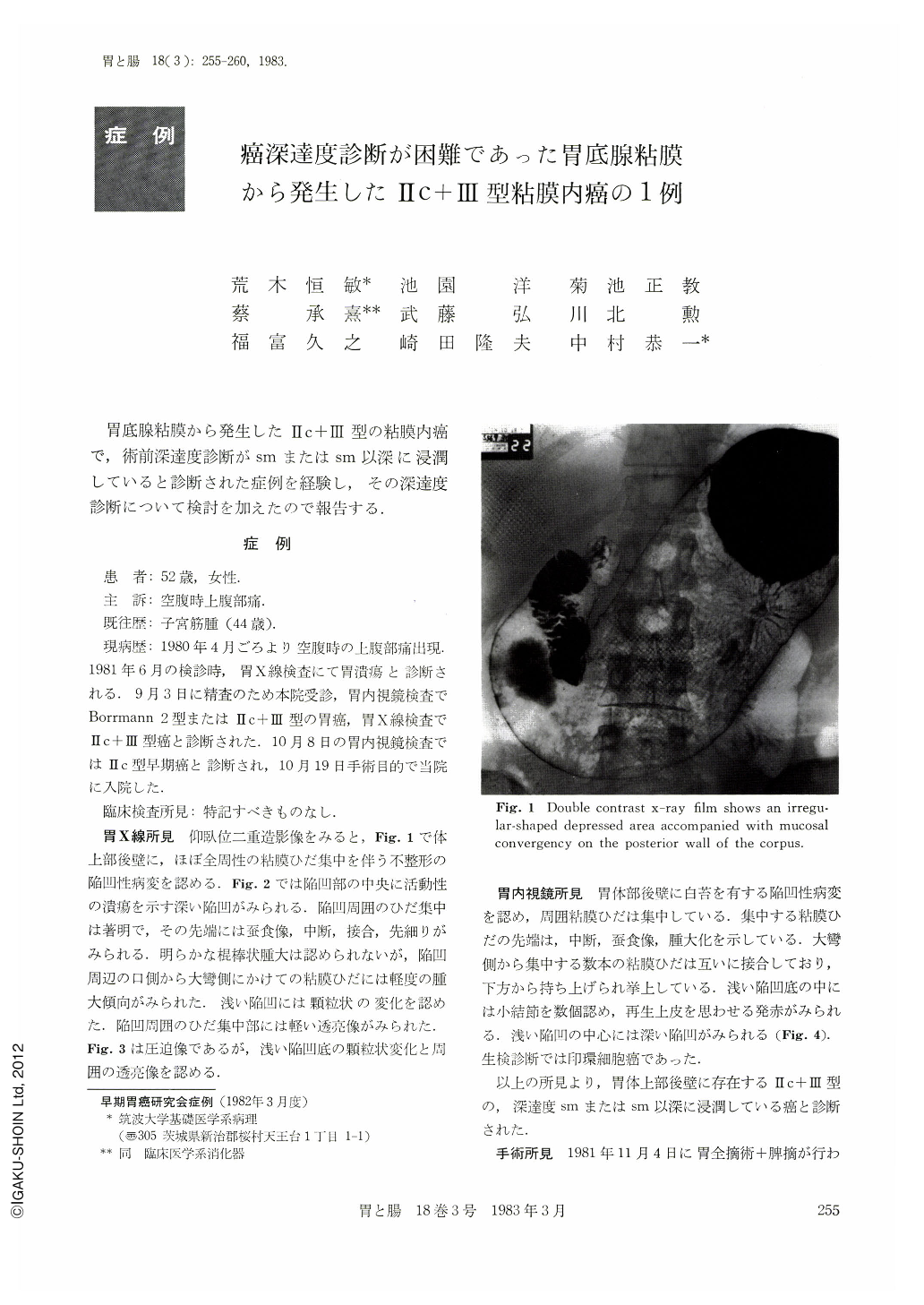
A 52-year-old woman had been suffering from epigastric pain before meals since April of 1980. In October of 1981, an x-ray examination of the upper gastrointestinal tract was performed at our hospital, and revealed a depressed lesion measuring about 3×2cm in size in the posterior wall of the gastric corpus, in the center of which a deep ulcer with converging mucosal folds was observed (Fig. 1). The converging mucosal folds showed abrupt interruption, swelling, and moth-eaten appearance (Fig. 2). Almost the same findings were seen by the following endoscopical examination. In addition, plateau-like elevation of the lesion was pointed out endoscopically (Fig. 4). The preoperative diagnosis made was an early cancer of Type Ⅱc+Ⅲ infiltrating the submucosa, or an adavnced cancer similar to Type Ⅱc+Ⅲ.
Total gastrectomy was performed on November 4, 1981 (Fig. 5). The depressed lesion of Type Ⅱc+Ⅲ measuring about 3×2cm in size is macroscopically located in the posterior wall of the corpus, in which there are many folds of the mucosa. A deep ulcer measuring about 1cm in diameter is observed in the center of the depressed lesion. Histological examination shows adenocarcinoma mucocellulare limited to the mucosa and completely situated in the fundic gland mucosa without intestinal metaplasia (Figs. 7, 9 and 10), and abundant submucosal fibrosis causing by ulceration. The area of submucosal fibrosis measures about 4.5×4.0cm in size (Figs. 6 and 7).
The submucosal fibrosis misled us to overestimate the depth of cancer invasion. In regard to radiological interpretation about vertical invasion of cancer with ulceration and situated in the area with many mucosal folds, it is very difficult to differentiate whether it is in the early phase or advanced. This is because submucosal fibrosis of ulceration in the area is more abundant than that in the antrum. It has to be considered that the radiological criteria for interpretation of vertical invasion of carcinoma situated in the area of many mucosal folds differs from that of carcinoma situated in the antrum.
Copyright © 1983, Igaku-Shoin Ltd. All rights reserved.