Japanese
English
- 有料閲覧
- Abstract 文献概要
- 1ページ目 Look Inside
- サイト内被引用 Cited by
最近の15年間にわれわれの教室で手術をうけた17例の非特異性小腸潰瘍患者のうち8例が組織学的に非特異性原発性多発性慢性小腸潰瘍と診断された(表1).このうちに2組の姉妹が含まれていたことは誠に興味ある点であったが,いずれにせよ他の4例と臨床的,組織学的にもほとんど差異を認めなかったので,この2組の姉妹の症例を述べ,本症に対するわれわれの考えをまとめてみたい.なおこれらの症例についてはすでに度々報告1)しているので症例報告とは少し異るが以下のようにまとめてみた.(これらの症例は本年の消化器病学会総会のシンポジウムで一部報告した)
症例
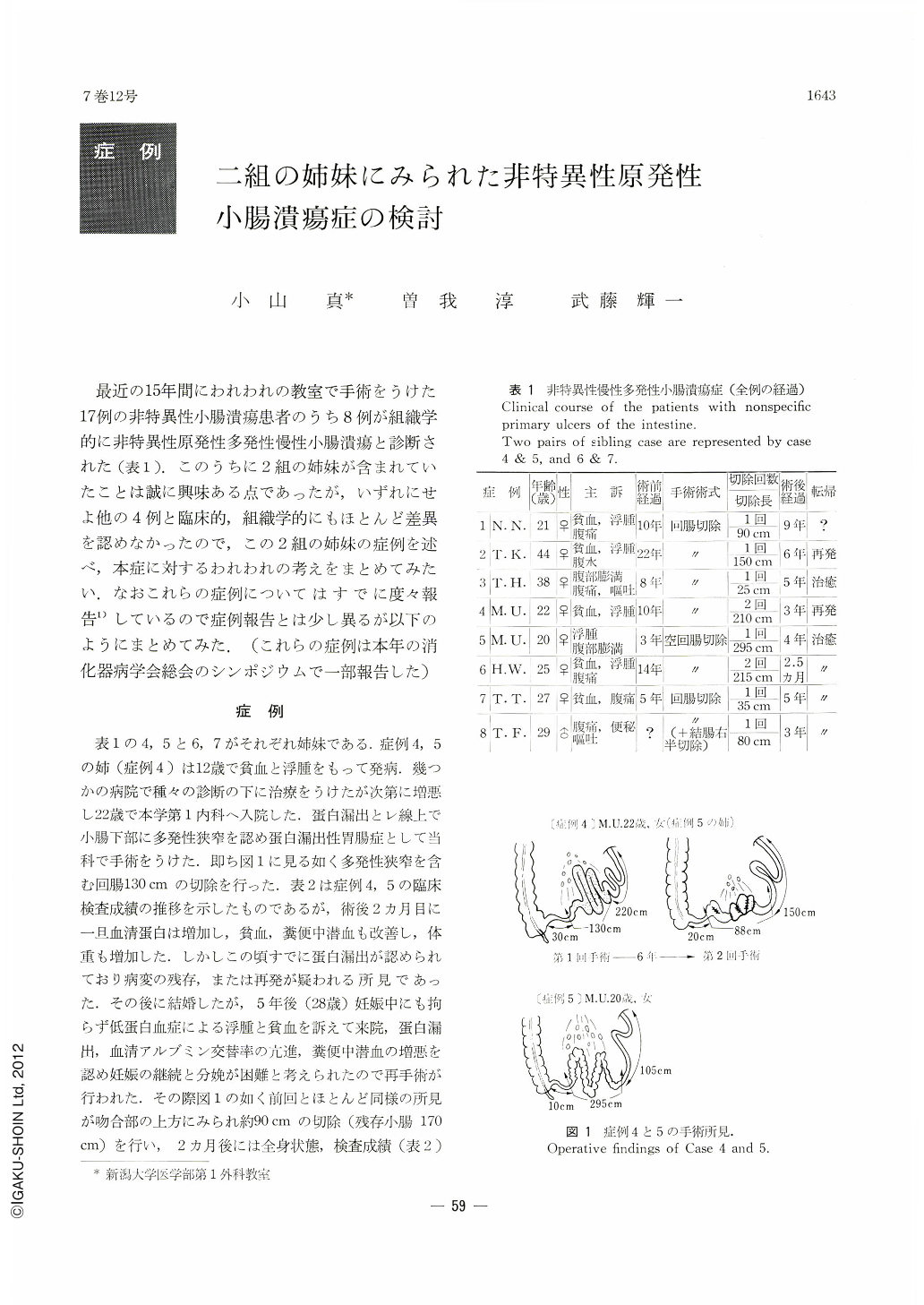
表1の4,5と6,7がそれぞれ姉妹である.症例4,5の姉(症例4)は12歳で貧血と浮腫をもって発病.幾つかの病院で種々の診断の下に治療をうけたが次第に増悪し22歳で本学第1内科へ入院した.蛋白漏出とレ線上で小腸下部に多発性狭窄を認め蛋白漏出性胃腸症として当科で手術をうけた.即ち図1に見る如く多発性狭窄を含む回腸130cmの切除を行った.表2は症例4,5の臨床検査成績の推移を示したものであるが,術後2カ月目に一旦血清蛋白は増加し,貧血,糞便中潜血も改善し,体重も増加した.しかしこの頃すでに蛋白漏出が認められており病変の残存,または再発が疑われる所見であった.その後に結婚したが,5年後(28歳)妊娠中にも拘らず低蛋白血症による浮腫と貧血を訴えて来院,蛋白漏出,血清アルブミン交替率の亢進,糞便中潜血の増悪を認め妊娠の継続と分娩が困難と考えられたので再手術が行われた.その際図1の如く前回とほとんど同様の所見が吻合部の上方にみられ約90cmの切除(残存小腸170
cm)を行い,2カ月後には全身状態,検査成績(表2)も改善し,その後無事出産することができた.しかし,2年後の昨年夏再び貧血,浮腫,無月経,下痢を主訴として来院,再発と診断され当科へ入院した.入院時の検査成績は表2,3に挙げてあるが,低蛋白血症,貧血と比較的軽度の蛋白漏出を認め,レ線でも本症再発が確認された.しかし,小腸広範囲切除によると考えられる蛋白,脂肪の吸収障害が認められたため,これ以上の腸管切除がはばかられたので輸血,輸血漿,栄養補液により全身状態の改善をみた.しかし輸血,輸液を中止すると間もなく検査所見の増悪をみ,更に吸収不良が認められたので試みに中鎖脂肪(MCT)の経口投与を行ってみたところ,幸いにもMC・8 250~300g/日(乳糖不耐症状を呈し3g/日のlactase剤の同時投与を要した)の経口投与で改善をみ約2週間後に退院せしめることができた.退院後今日まで200~250g/日のMC・8と9錠/日のサラゾピリン,2錠/日のフェログラジュメットの経口投与で一応良好に経過している.
Clinical course: The onset of the patients' symptoms was at a relatively young age of 11 to 22, and after a long period (3 to 14 years) of anemia, edema, diarrhoea, occult blood in the stool and, in some cases, intermittent crampy abdominal pain, were they diagnosed as having “proteinlosing gastroenteropathy”. They all showed severe hypoproteinemia and increased fecal excretion of albumin on Gordon's test. A G-I series exhibited multiple areas of stenosis in the small intestine suggesting ulcer formation with dilatation of the bowel proximal to the sites of stenosis. At the time of resection of the diseased bowels (ranging from 35 to 295 cm in length), multiple areas of annular stricture and ulceration with enlarged soft mesenteric lymph nodes were observed.
Two patients, the elder sisters of each of two siblings, received re-operation at 6 and 5 years respectively, after the first operation; the intestine resected showed the same type of the disease. While one of these 2 patients has been free of the disease since re-operation, the other has been suffering from recurrent episodes of the disease. The immunotolerance of the latter patient was confirmed by a long time survival (at least for 45 days) of a homotransplanted skin flap. For the last 2 months, she has received a bulk of medium chain triglyceride (MCT) (about 40~60 gm/day) and slazopyrin (9 Tab/day) orally, but, fortunately, she has maintained a relatively healthy state.
Pathologic examination: Upon opening the resected intestine, we detected striking abnormalities found in common as follows: (1) multiple superficial ulcers with narrowing of the intestinal lumen, (2) skip areas of relatively normal mucosa, and (3) in most cases no remarkable thickening of the bowel wall. Two to 20 annular ulcers were observed in all cases. Microscopically, the ulcers were mostly superficial, extending only to the muscularis mucosae or submucosa associated with underlying chronic inflammatory changes. The submucosa, under or around the ulcers, was slightly edematous and replaced by marked fibrosis with moderate infiltration of plasma cells, lymphocytes and fewer eosinophils. Secondary proliferation of lymphoid follicles was seen in the mucosa and submucosa in two cases, but marked dilatation of lymphatics of the bowel wall and lymph nodes was not observed in them. None exhibited granulomas supposedly characteristic of Crohn's disease.
Conclusion: 1) Since, in some cases, medical treatment such as oral administration of MCT and antibiotics is effective, this should be attempted on all patient suspected of having primary ulcers of the small intestine. 2) As in the cases of regional enteritis, the extent of resection of the intestine should be 20 to 30 cm proximal to the limit of the visible lesions because of a high incidence of recurrence in the areas immediately proximal to site of anastomosis.
Copyright © 1972, Igaku-Shoin Ltd. All rights reserved.