Japanese
English
- 有料閲覧
- Abstract 文献概要
- 1ページ目 Look Inside
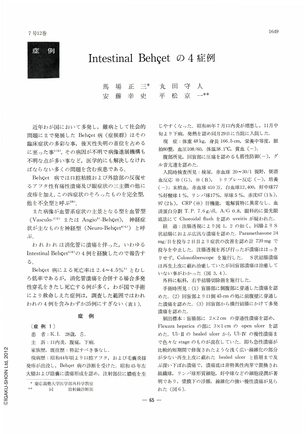
近年わが国において多発し,難病として社会的問題にまで発展したBehçet病(症候群)はその臨床症状の多彩な事,後天性失明の首位を占めるに至った事1)2),その病因が不明で病像進展機構も不明な点が多い事など,医学的にも解決しなければならない多くの問題を含む疾患である.
Behçet病では口腔粘膜および外陰部の反復せるアフタ性有痛性潰瘍及び眼症状の三主徴の他に皮疹を加え,この四症状のそろったものを完全型,他を不全型と呼ぶ29).
In Japan the incidence of Behçet's disease has been increasing significantly and now it has become a social problem. In some cases it is accompanied with gastrointestinal symptoms, so that it is then called intestinal Behçet or gastrointestinal Behçet disease.
Ulcers seen in this disease tend to perforate. Four cases of intertinal Behcet disease, treated in our University Hospital, were either perforated or penetrated. Including a patient with panperitonitis, all were rescued and had uneventful postoperative courses. In one patient, clinical remission of other mucocutaneous ocular symptoms was observed after the operation.
Reviewing the Japanese literature, we have collected 25 survival cases by successful operation, including our 4 cases. Analysis of 25 cases shows that out of 17 penetrated or perforated ulcers, 7 were multiple perforations. Ulcers were mostly located in the terminal ileum or in the right half of the colon. There is a difference in the locality of the lesion between ulcerative colitis and intestinal Behçet disease.
As most of perforations occurred in the right half of the colon, right hemicolectomy with end-to-end anastomosis seems to be an operation of choice. It is advisable to resect the ileum approximately 50 cm long from the ileocecal valve in order to prevent recurrence of ulcers.
Characteristic fluoroscopic pictures of the colon showing en face or profile niche in intestinal Behçet disease are demonstrated here. Ulcers in this disease tend to undermine superficial layers, being deep enough to reach into the muscular or serosal layer. Diagnosis was established preopearatively in 2 out of 4 treated cases. Ulcers were observed colonoscopically and it is considered to be the first case in the world in which ulcers due to intestinal Behçet disease were directly observed by colonoscopy.
Microangiography was performed to the resected specimen, revealing disturbed microcirculation surrounding the ulcers especially in the submucosal vessels.

Copyright © 1972, Igaku-Shoin Ltd. All rights reserved.


