Japanese
English
- 有料閲覧
- Abstract 文献概要
- 1ページ目 Look Inside
人間ドックによる健康診断で,たまたま無症状で発見され,根治手術可能であった多中心性に発生したと思われる粘膜内胆管癌の1例を経験したので,文献的考察を加え報告する.
症例
患 者:K.T.,60歳,女.
主 訴:特別訴えるような自覚症状はない.
既往歴:45歳のとき,卵巣嚢腫摘出術および虫垂切除術を受ける.
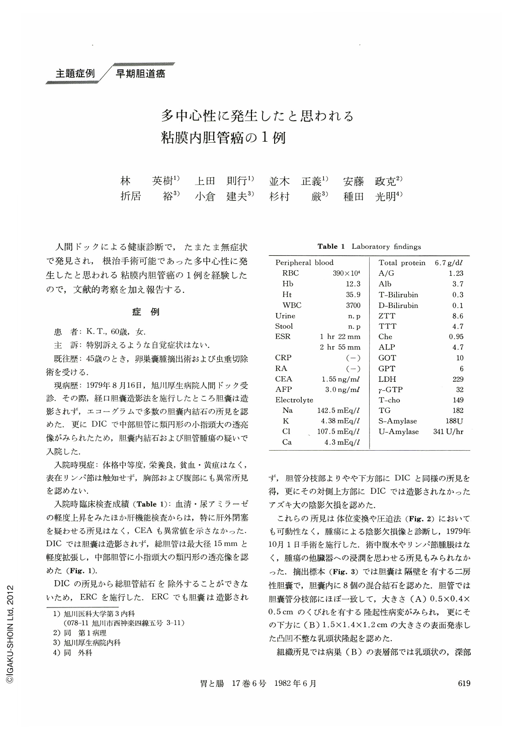
現病歴:1979年8月16日,旭川厚生病院人間ドック受診.その際,経口胆嚢造影法を施行したところ胆嚢は造影されず,エコーグラムで多数の胆嚢内結石の所見を認めた.更にDICで中部胆管に類円形の小指頭大の透亮像がみられたため,胆嚢内結石および胆管腫瘍の疑いで入院した.
This is a case of a woman aged 60 years in whom gallstones were diagnosed. Echogram revealed multiple stones in the gallbladder although the gallbladder could not be visualized with oral cholangiography on a routine health examination. DIC revealed a mildly dilated choledochus with a maximum diameter of 15 mm and a spherical filling defect about the size of the tip of the little finger in the middle portion of the common bile duct. ERC was performed to rule out any possible choledocholithiasis. This resulted in negative visualization of the gallbladder and the same finding as with DIC for the common bile duct.
Furthermore, a filling defect about the size of a small green pea was recognized on the upper opposite site. This was not shown with DIC.
The extirpated gallbladder had two atria divided by a septum which contained 8 mixed cholesterol stones. There was a protruded lesion of 0.5×0.4×0.5 cm with a stalk at the site of the branching of the cystic duct from the common bile duct. In the duodenal side of the common bile duct, there was another 1.5×1.4×1.2 cm papillary lesion, which had an irregular surface manifesting hyperemic reddening. The histology of their lesions documented papillary adenocarcinoma which was intramucosal carcinoma (carcinoma in situ). These two lesions were separated by normal mucosa; thus multicentric carcinoma in situ seemed to be the most probable diagnosis.
Copyright © 1982, Igaku-Shoin Ltd. All rights reserved.