Japanese
English
- 有料閲覧
- Abstract 文献概要
- 1ページ目 Look Inside
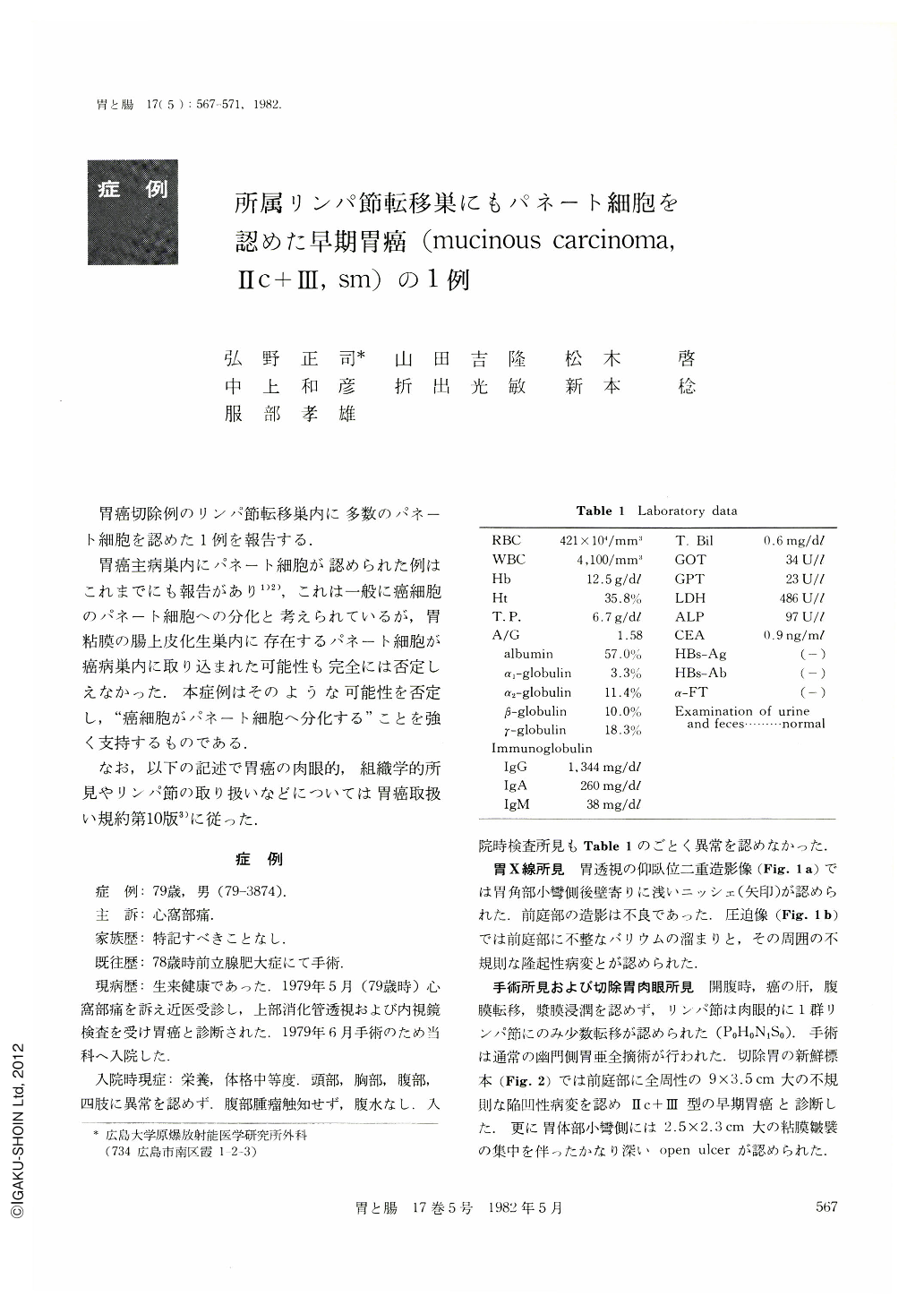
胃癌切除例のリンパ節転移巣内に多数のパネート細胞を認めた1例を報告する.
胃癌主病巣内にパネート細胞が認められた例はこれまでにも報告があり1)2),これは一般に癌細胞のバネート細胞への分化と考えられているが,胃粘膜の腸上皮化生巣内に存在するパネート細胞が癌病巣内に取り込まれた可能性も完全には否定しえなかった.本症例はそのような可能性を否定し,“癌細胞がパネート細胞へ分化する”ことを強く支持するものである.
The resected specimen from this 79-year-old man showed a depressed, Ⅱc+Ⅲ type early gastric cancer in the antrum and a round open ulcer in the body. Histology revealed mucinous carcinoma with its invasion limited to the submucosal layer and an UI-Ⅳ open ulcer. Two regional lymph nodes in the lesser omentum of the stomach were positive for metastatic carcinoma. Paneth cells were found in a part of the primary lesion and, to be noteworthy, in the two metastasized lymph nodes. Enzyme-labeled antibody method was performed for the detection of an enzyme lysozyme, which is commonly considered to be characteristic of Paneth cells.
Few cases of gastric carcinoma have so far been reported in which Paneth cells are found in the primary lesion. However, Paneth cells are usually present in intestinalized mucosa of the stomach and it must be strictly ruled out that these benign Paneth cells are involved in carcinoma tissue in the course of carcinoma growth. This case strongly suggests that Paneth cells in carcinoma tissue have originated, not in intestinalized mucosa, but in carcinoma cells.

Copyright © 1982, Igaku-Shoin Ltd. All rights reserved.


