Japanese
English
- 有料閲覧
- Abstract 文献概要
- 1ページ目 Look Inside
- サイト内被引用 Cited by
食道静脈瘤の内視鏡診断は,食道ファイバースコープの開発されなかった硬性食道鏡の時代では,ほとんど行われなかったのが実情であった.食道静脈瘤は食道鏡の禁忌とするものも,また検査を行いうるとしたものもあるが1),十分な観察は困難であったようで,詳細な観察所見の発表は少なかった.
わが国において食道ファイバースコープの導入が行われたのは,著者らがアメリカンチストスコープ社製食道ファイバースコープを用いて,その結果を1965年春,第7回内視鏡学会に発表したのが最初であった2).
Esophageal varices are classified into 3 grades on the ground of fiberscopic examinations performed on 117 patients, with 263 repeated examinations.
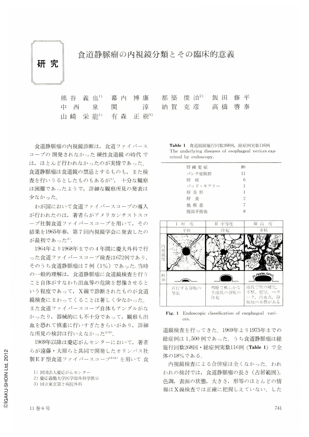
Grade 1 is characterized by dilated submucosal veins, which do not bulge into the esophageal lumen.
Grade 2 is observed as semispherical protuberance of submucosal varices with smooth surface into the esophageal lumen.
Grade 3 is presented as protuberance of dilated tortuous varices with superimposed varices into the esophageal lumen, which are similar to varices on top of varices designated by Dagradi, close correlation observed between the frequency of bleeding and the grade of the esophageal varices. None bled in grade 1 and 6 out of 29 patients classified into grade 2 experienced hemorrhage, indicating the frequency of 20 percent, while 32 out of 50 patients of grade 3 bled with a frequency of 64 percent. There were 6 bleeding cases classified into Grade 2 ; 2 were due to gastric bleeding, the other 2 had esophageal erosion due to reflux esophagitis and 1 had marked ascites, which also tends to cause reflux esophagitis.
These observations led as to conclude that patients of grade 3 are very liable to bleed and they should be undergone esophageal transection even if they have not experienced hemorrhage before. And it is also suggested that grade 2 varices with reflex esophagitis is an indication for esophageal transection, since reflex esophagitis is a precipitating factor in variceal bleeding.

Copyright © 1976, Igaku-Shoin Ltd. All rights reserved.


