Japanese
English
- 有料閲覧
- Abstract 文献概要
- 1ページ目 Look Inside
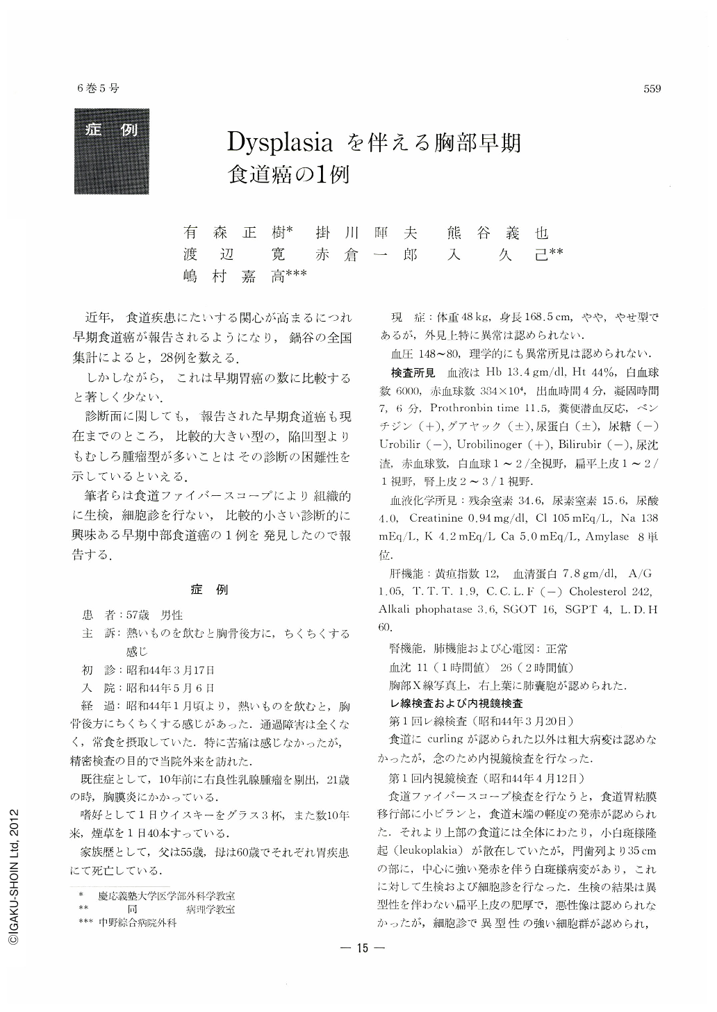
近年,食道疾患にたいする関心が高まるにつれ早期食道癌が報告されるようになり,鍋谷の全国集計によると,28例を数える.
しかしながら,これは早期胃癌の数に比較すると著しく少ない.
58-year-old male who complained of moderate retrosternal pain was examined by x-ray examination and esophagoscopy performed simultaneously with biopsy and cytology in the Keio University Hospital.
At 35 cm from the incisor teeth, a small erosion was seen by esophagofiberscopic observation. Biopsy from this lesion revealed squamous cell carcinoma. Subtotal esophagectomy and retrosternal esophagogastrostomy was performed. No swelling of the lymphatic glancl was recoginized at the time of operation. Cancerous lesion was 1.0×0.9 cm and cancer was confined within the submucosal layer and did not infiltrate into the muscle layer. The surface of cancer lesion was exposed over the mucosa in 1.7×0.2cm and around the erosion the mucosa was protruded slightly due to the submucosal infiltration of cancer cells. There was no metastasis of the lymphatic gland microscopically.
Around this cancerous lesion there were many scattered epithelial dysplasia in various grade. Endoscopically, it was very difficult to differentiate cancerous lesion from these small epithelial dysplasia.
Erosion around leucoplakia-like dysplasia has to be carefully checked by biopsy or cytology. Cancerous lesion seems to be more reddish in comparison with these epithelial dysplasia endoscopically.
Reproducibility of this small cancer by X-ray examination is diflicult and esophagofiberscopic examination is very useful for detection of these small cancer lesions.

Copyright © 1971, Igaku-Shoin Ltd. All rights reserved.


