Japanese
English
- 有料閲覧
- Abstract 文献概要
- 1ページ目 Look Inside
日本消化器病学会クローン病検討委員会からクローン病の診断基準(案)1)が提出され,今迄かなり混乱していた本疾患がひとつのdisease entityとして検討できるようになった,しかし,病変の進行度にもよるのであるが,肉眼形態の上でかなりの差異を認める症例がある.われわれも最近,非定型的症例を経験したので報告する.
症例
患 者:M. K. 29歳 男性 公務員
主 訴:右下腹部痛
家族歴:特記すべきことなし
既往歴:1972年12月,虫垂炎にて切除術を受けた.
現症歴:上記虫垂切除術を受けた後,右下腹部に軽度の圧痛が続いていた.1975年10月同部に激痛が現れたが,1日で消退したため放置していた.1976年6月再び右下腹部に激痛があり,近医受診,触診にて同部にクルミ大の腫瘤を指摘され,注腸検査により回盲部に狭窄を認め,大学病院に紹介された.この間に,下痢,下血なく,また悪心,嘔吐などの消化器症状も認めていない.発熱なし.
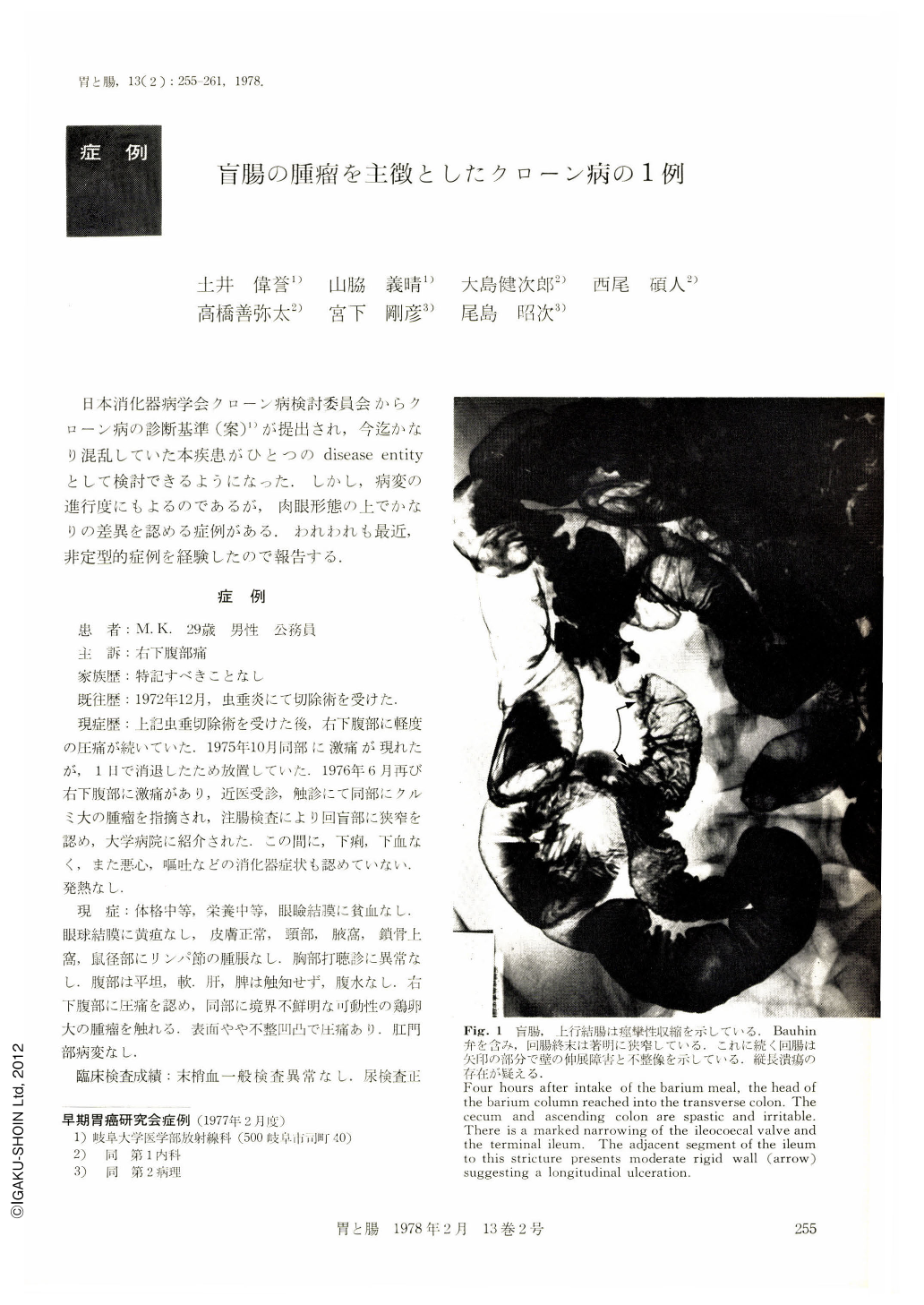
The patient is a 29-year-old male. In December 1972, he had undergone appendectomy. Since then he frequently complained of some discomfort with mild tenderness at the right lower abdomen. In June 1976, he suffered from severe colicky pain at this region without any onset of melenea or diarrhoea. Alimentery tract X-ray examination revealed marked spastic shrinkage of the cecum and ascending colon. The Bauhin's valve and terminal ileum presented marked stricture, but there was no evidence of obstruction. Barium enema and colonofiberscopy revealed a bilocular elevated lesion at the bottom of the coecum suggesting a submucosal tumor. The surface of the tumor appeared smooth with no evidence of mucosal effacement. There was no evidence of a deep ulceration or of a fistula formation. The terminal ileum involving the ileocecal valve presented a marked narrowing, and the proximal segment adjacent to this stricture was consequently dilated with an irregular unilateral rigidity of the wall.
In July 1976, partial resection from the distal segment of the ileum to the proximal ascending colon was performed. Small amount of serous ascites was found. There was tight adhesion between the cecum and the terminal ileum. No fistula formation was noted. On the resected specimen, a longitudinal ulcer was found at the terminal ileum. Several small erosions and ulcerations were seen on the region of the ileocoecal valve. Histological study disclosed that transmural inflammation spreads across all layers of the coecum and several fistulas reaches into the mucosa and submucosa. There are small abscesses surrounded by non-caseating epitheloid cell granulation observed in the central portion of the submucosal layers. Regional mesenteric lymph nodes also present non-caseating sarcoid-like granuloma with a few Langhans' giant cells.
According to the diagnostic criteria of Crohn's disease proposed by the Japanese Society of Gastroenterology, this case may be adopted to the category of this disease, eventhough the clinical features are not enough significant. It is understandable that the clinical features may vary depending on the stage of progress or on individual difference of immunological reaction.

Copyright © 1978, Igaku-Shoin Ltd. All rights reserved.


