Japanese
English
- 有料閲覧
- Abstract 文献概要
- 1ページ目 Look Inside
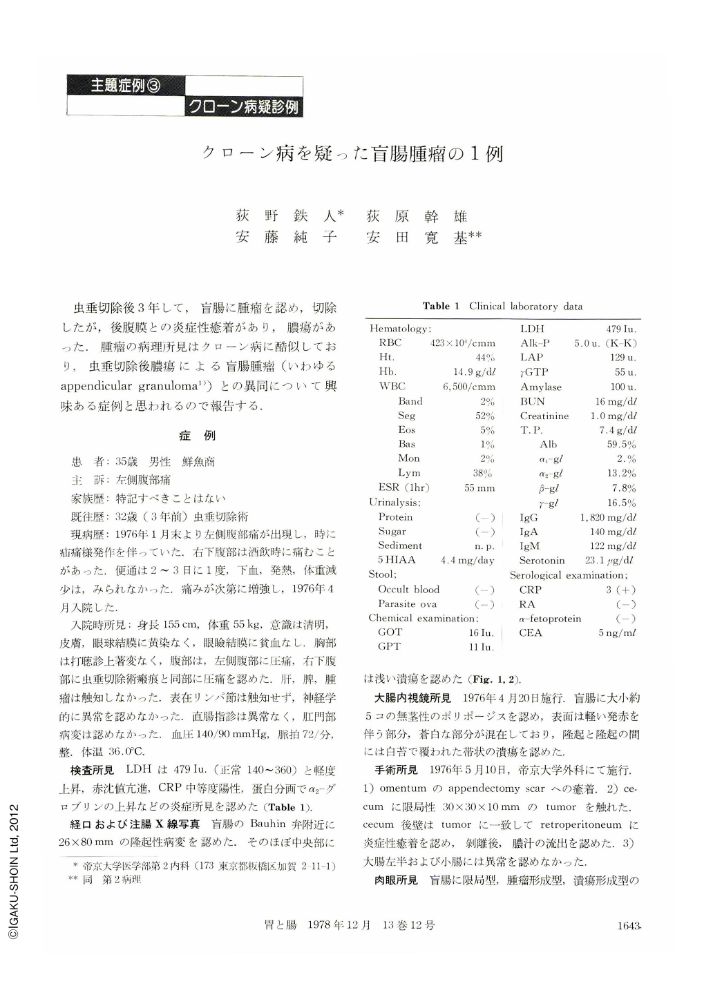
虫垂切除後3年して,盲腸に腫瘤を認め,切除したが,後腹膜との炎症性癒着があり,膿瘍があった.腫瘤の病理所見はクローン病に酷似しており,虫垂切除後膿瘍による盲腸腫瘤(いわゆるappendicular granuloma1))との異同について興味ある症例と思われるので報告する.
A 35-year-old man was admitted to the hospital because of left side abdominal pain. He was well until three years earlier, when appendectomy was performed. Two and a half months before admission, he began to experience a sharp pain in the left side of the abdomen. Subsequently pain developed sometimes in the right lower quadrant of the abdomen especially after drinking alcohol. There was no change in weight or history of melena and fever.
Examination showed the following. The temperature 36.0℃, the pulse 72/min, blood pressure was 140/90 mmHg. Abdominal examination disclosed normal bowel sound with tenderness in the left side and in the right lower quadrant where appendectomy scar was seen; no organ, mass or spasm was felt. No lymphadenopathy was found. Digital examination was negative, without perianal lesions. The hematocrit was 44%; the white cell count 6,500 with normal differential count. The erythrocyte sedimentation rate was 55 mm per hour. The protein was 7.4 g per 100 ml (the albumin 59.5%, α1-gl 2.5%, α2-gl 13.2%, β-gl 7.8%, γ-gl 16.5%), the lactic dehydrogenase (LDH) was 479 Iu (normal 140~360). C reactive protein was three plus positive. A stool specimen gave a negative test for occult blood and parasite ova. Barium enema revealed lesions or filling defect in the cecum (26×80 mm) with shallow ulcer and finally colonofiberscope disclosed five polypoid lesions with ulcers.
Operation was performed on May 10, 1976. Findings were as follows. 1) Adhesion of omentum to appendectomy scar. 2) Retrocecum was adherent inflammatorily to retroperitoneum with dischage of pus. Macroscopically, there were five polypoid lesions (ca 10×20 mm) without stiel, with shallow ulcer.
Microscopically, the basal portion of ulcer shows granulomatous lesion with abundunt capillary vascular elements and extensive round cell infiltration. Subserosal granulomatous lesion fibroreticulosis with abundant vascular elements and several giant cells.
Conclusively the cecal tumor following perityphilic abscess, tuberculosis, or sarcoidosis, should be ruled out, but, these clincal and pathological findings are suggestiveof so-called Crohn's disease.

Copyright © 1978, Igaku-Shoin Ltd. All rights reserved.


