Japanese
English
- 有料閲覧
- Abstract 文献概要
- 1ページ目 Look Inside
近年,胃X線,内視鏡検査および生検の進歩とともに,胃原発悪性リンパ腫の症例は数多く報告されている.しかし,臨床的には良性悪性との鑑別診断が問題となり,他方,病理組織学的にも限局性のリンパ細網細胞増生との鑑別診断が問題となる.われわれは胃悪性リンパ腫と診断,手術し,術後も悪性と考えて化学療法を施行したが,病理組織学的には良性悪性の鑑別が困難であった1症例を経験したので報告する.
症例
患 者:57歳 女性 主婦
主 訴:空腹時の上腹部痛
既往歴:1976年9月急性虫垂炎で虫垂切除
家族歴:祖父が胃癌で死亡以外,特記すべきことなし.
現病歴:約5年前より胸やけ,背部痛および曖気があって,某医を受診,慢性胃炎の診断で治療を受けていた.過去1年間に約20kgの体重減少があり,空腹時に嘔気と上腹部痛を訴えるようになり,精査のため1975年12月1日本院に入院した.
The patient, a 57-year-old housewife, was admitted to our hospital on Dec. 1, 1975, because of hunger pain in the epigastrium and weight loss of 20 kg for the past one year.
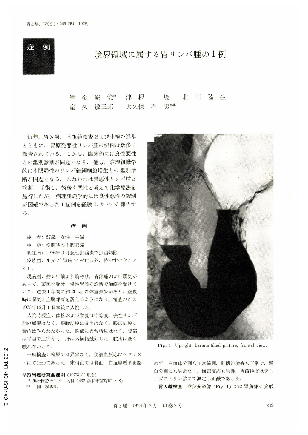
The upper GI series showed numerous small and shallow ulcerations, accompanied with converging mucosal folds of which tips were enlarged. These ulcerations were widely spread from the infracardiac portion to the antrum on both aspects of anterior and posterior walls. The walls expanded well.
Endoscopic examination showed in the same place multiple erosions and ulcerations accompanied with thickened, converging mucosal folds, and tendency to bleed easily. The biopsy and cytology examinations revealed findings suggestive of malignant lymphoma. Under a diagnosis of malignant lymphoma, total gastrectomy was performed. No swelling of resional lymphnodes was noted. In the resected specimen, multiple ulcers and erosions were noticed in the same area as we had expected preoperatively.
The tips of the converging fold were enlarged and confluent and partially looked like bridging folds suggestive of a submucosal tumor.
Histologically,12 ulcers were observed, all Ul-Ⅱ type of Murakami's classification. Lymphoid cells and reticulum cells were proliferating in the center of each ulcer independently or confluently and invading only the adjacent submucosal layer. As the histological characteristics of lymphoid tissue, the following findings were observed : in part lymphoreticular hyperplasia which had enlarged lymph follicles with germinal centers; in part diffuse proliferation of immature lymphocytes which was suggestive of malignant lymphoma; and in part intermingled features of the above. From these findings, this case was diagnosed as lymphoma which belonged to the border line region.

Copyright © 1978, Igaku-Shoin Ltd. All rights reserved.


