Japanese
English
- 有料閲覧
- Abstract 文献概要
- 1ページ目 Look Inside
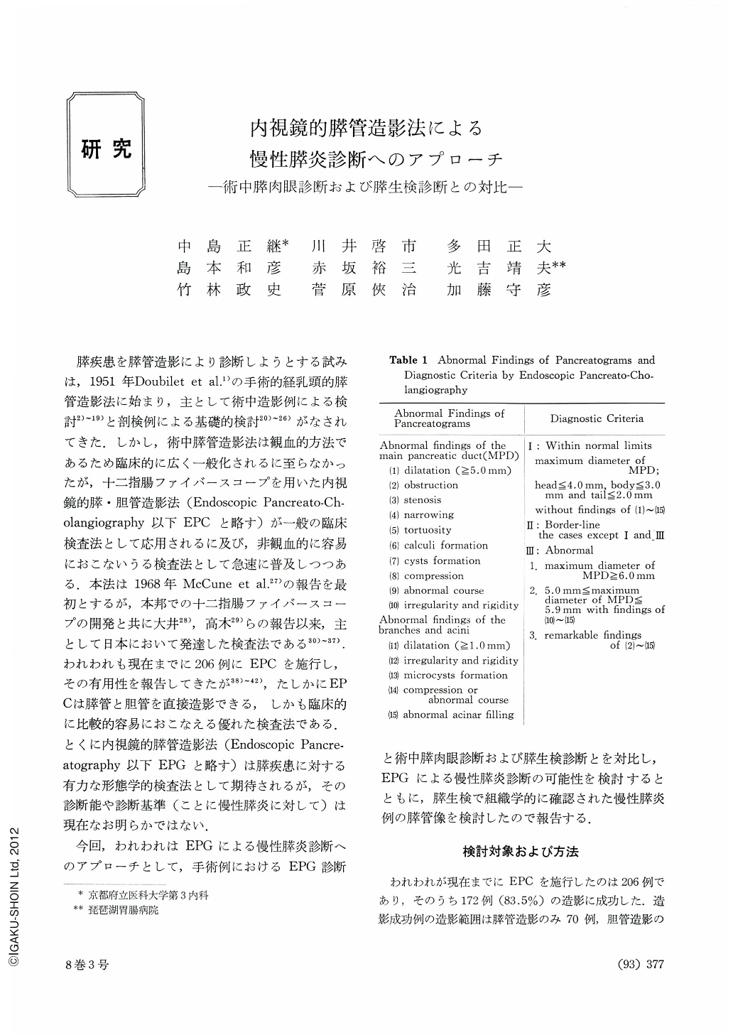
膵疾患を膵管造影により診断しようとする試みは,1951年Doubilet et al.1)の手術的経乳頭的膵管造影法に始まり,主として術中造影例による検討2)~19)と剖検例による基礎的検討20)~26)がなされてきた.しかし,術中膵管造影法は観血的方法であるため臨床的に広く一般化されるに至らなかったが,十二指腸ファイバースコープを用いた内視鏡的膵・胆管造影法(Endoscopic Pancreato-Cholangiography,以下EPCと略す)が一般の臨床検査法として応用されるに及び,非観血的に容易におこないうる検査法として急速に普及しつつある.本法は1968年McCune et al.27)の報告を最初とするが,本邦での十二指腸ファイバースコープの開発と共に大井28),高木29)らの報告以来,主として日本において発達した検査法である30)~37).われわれも現在までに206例にEPCを施行し,その有用性を報告してきたが38)~42),たしかにEPCは膵管と胆管を直接造影できる,しかも臨床的に比較的容易におこなえる優れた検査法である.とくに内視鏡的膵管造影法(Endoscopic Pancreatography,以下EPGと略す)は膵疾患に対する
有力な形態学的検査法として期待されるが,その診断能や診断基準(ことに慢性膵炎に対して)は現在なお明らかではない.
今回,われわれはEPGによる慢性膵炎診断へのアプローチとして,手術例におけるEPG診断と術中膵肉眼診断および膵生検診断とを対比し,EPGによる慢性膵炎診断の可能性を検討するとともに,膵生検で組織学的に確認された慢性膵炎例の膵管像を検討したので報告する.
To establish the role of endoscopic pancreatography as a diagnositic approach to chronic pancreatitis, we have positively employed biopsy during surgical exploration in cases whose pancreas had been preoperatively visualized. Aside from biopsied cases of cancer of the pancreas, its cyst and its secondary infiltration with gastric cancer, 35 independent cases were examined for correlating their pancreatographic pictures with gross findings including those of palpation and with histologic dianosis of biopsied tissue. Attempts have also been made to make sure diagnostic possibility of endoscopic procedure in chronic pancreatitis, together with a study of pictures of the ducts in chronic inflammation as confirmed by biopsy.
1. Of 35 cases of well opacified pancreatic ducts, 16 were determined as within normal limits, 5 on a borderline between the normal and abnormal and 14 as abnormal (suspect of chronic pancreatitis). On the other hand, gross findings in 23 cases were judged as within normal limits and those of the remaining 12 were interpreted as cirrhotic (suspect of chronic inflammation). In histodiagnosis of biopsied tissue 22 cases were normal, 2 were of fatty degeneration, 1 of atrophy and 10 of chronic pancreatitis.
2. A great deal of similarity was seen between the results of pancreatography and gross findings or histodiagnosis of biopsied materials. Special care has been taken to compare the results of biopsy and pancreatography at the sites as close as possible to each other, and yet 9 out of 10 cases diagnosed by biopsy as chronic pancreatitis were also judged by pancreatography as showing abnormal findings in the ducts. The more abnormal the ducts in opacified pictures were, the more so were their gross findings. The same tendency was seen histologically as well.
3. Pancreatographs in 10 cases histologically diagnosed as chronic inflammation showed dilataion and rigidity of the main duct along with irregular stiffness and small cyst formation in the branching ducts. In highly advanced cases, even narrowing and twisting of the main duct was seen together with dilatation of the branching ducts. When inflammation was not so severe, the main duct displayed no particular changes, most cases showing only irregular rigidity or cyst formation in the branching ducts or else partial acinar abnormality.

Copyright © 1973, Igaku-Shoin Ltd. All rights reserved.


