Japanese
English
- 有料閲覧
- Abstract 文献概要
- 1ページ目 Look Inside
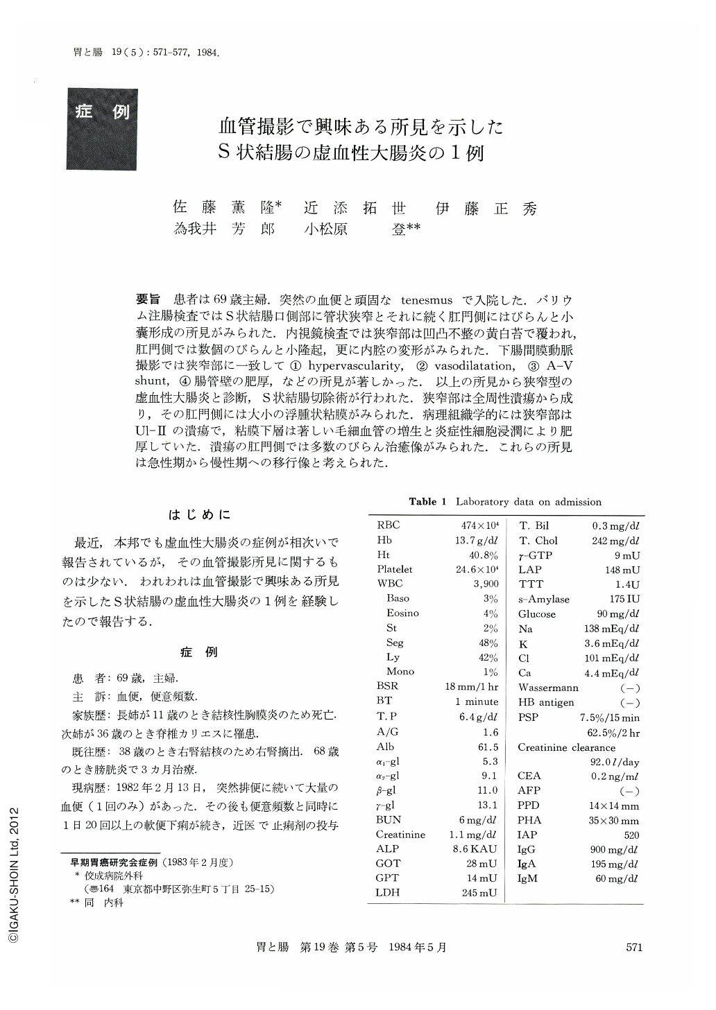
要旨 患者は69歳主婦.突然の血便と頑固なtenesmusで入院した.バリウム注腸検査ではS状結腸口側部に管状狭窄とそれに続く肛門側にはびらんと小嚢形成の所見がみられた.内視鏡検査では狭窄部は凹凸不整の黄白苔で覆われ,肛門側では数個のびらんと小隆起,更に内腔の変形がみられた.下腸間膜動脈撮影では狭窄部に一致して①hypervascularity,②vasodilatation,③A-Vshunt,④腸管壁の肥厚,などの所見が著しかった.以上の所見から狭窄型の虚血性大腸炎と診断,S状結腸切除術が行われた.狭窄部は全周性潰瘍から成り,その肛門側には大小の浮腫状粘膜がみられた.病理組織学的には狭窄部はUl-Ⅱの潰瘍で,粘膜下層は著しい毛細血管の増生と炎症性細胞浸潤により肥厚していた.潰瘍の肛門側では多数のびらん治癒像がみられた.これらの所見は急性期から慢性期への移行像と考えられた.
The patient, a housewife aged 69, was admitted to the hospital because she had melena, although once, and persistent tenesmus. On the 33rd day after admission barium enema revealed on the oral side of the sigmoid a tubular narrowing, 9 cm long and 1.0 cm wide, and erosions and sacculation adjacent to the above on the anal side of the sigmoid. Biopsy of the same place showed uneven. yellowish white coating in the narrowed part and several erosions, small elevations and intraluminal deformity in the adjacent anal side of the sigmoid. Angiography of the inferior mesenteric artery showed such striking findings at the site corresponding to the tubular narrowing as 1) hypervascularity, 2) vasodilatation, 3) A-V shunt, 4) thickened intestinal wall and so forth. These findings led us to a diagnosis of stricturing type of ischemic colitis. On the 55 th day (April 9, 1982) after the onset of the disease sigmoidectomy was performed, The stricture was caused by a circumferential ulcer 8 cm long. On its anal side was seen edematous mucosa of various size. Histologically the narrowing was caused by a Ul-II ulcer. The thickened submucosa showed striking proliferation of capillary veins and infiltration of inflammatory cells. On the anal side of the ulcer were seen erosions in various healing stages. These findings were considered as those of transitional stage of ulcer from acute to chronic one.

Copyright © 1984, Igaku-Shoin Ltd. All rights reserved.


