Japanese
English
- 有料閲覧
- Abstract 文献概要
- 1ページ目 Look Inside
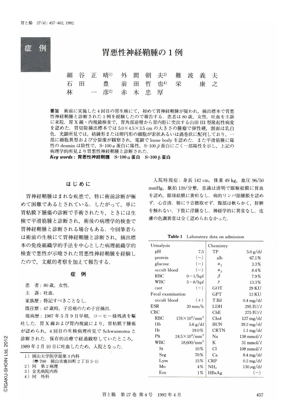
要旨 術前に実施した4回目の胃生検にて,初めて胃神経鞘腫が疑われ,摘出標本で胃悪性神経鞘腫と診断された1例を経験したので報告する.患者は80歳,女性.吐血を主訴に来院.胃X線・内視鏡検査で,胃角部前壁から胃内腔に突出する山田Ⅲ型隆起性病変を認めた.胃切除摘出標本では5.0×4.5×3.5cmの大きさの腫瘤で弾性硬,割面は乳白色.光顕所見では,紡錘形または卵円形の細胞が索状あるいは渦巻状に配列しており,一部に細胞異型および分裂像が観察され,電顕でloose bodyを認めた.また平滑筋腫に陽性のdesminは陰性で,S-100α蛋白に陽性,S-100β蛋白にごく一部陽性を示し,上記の病理学的所見より胃悪性神経鞘腫と診断された.
An 80-year-old female visited our hospital complaining of vomiting of blood. Endoscopic examination and radiography showed a submucosal tumor, 5×4.5×3.5 cm in size, in the anterior wall of the angulus. It was classified as Yamada's type Ⅲ. The tumor, protruding into the lumen of the stomach, was elastic firm, well circumscribed and whitish and partly gray on cut surface. Light microscopic examination revealed a densely cellulated tumor consisting mostly of spindle-shaped cells. There were partly pleomorphism and only a few mitoses (Fig.5 c). Electron microscopy showed spindle cells and long-spacing collagen (loose body). Immunohistochemically, S-100 and S-100 α were positive but desmin negative. S-100β was only slightly positive.
Malignant Schwannoma of the stomach is very rare. It is difficult to differentiate it from leiomyosarcoma not only clinically but pathologically in most cases. Immunohistochemical staining for S-100 α, S-100 β and desmin is useful in histologically differentiating neurogenic sarcoma from myogenic tumors.

Copyright © 1992, Igaku-Shoin Ltd. All rights reserved.


