Japanese
English
- 有料閲覧
- Abstract 文献概要
- 1ページ目 Look Inside
- サイト内被引用 Cited by
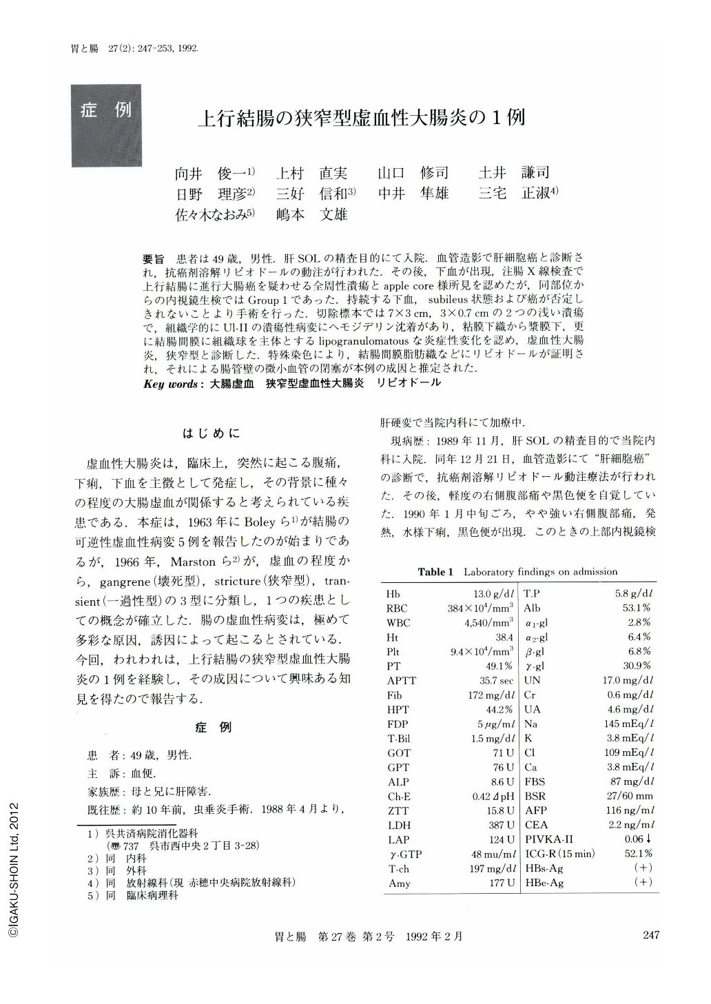
要旨 患者は49歳,男性.肝SOLの精査目的にて入院.血管造影で肝細胞癌と診断され,抗癌剤溶解リピオドールの動注が行われた.その後,下血が出現,注腸X線検査で上行結腸に進行大腸癌を疑わせる全周性潰瘍とapple core様所見を認めたが,同部位からの内視鏡生検ではGroup 1であった.持続する下血,subileus状態および癌が否定しきれないことより手術を行った.切除標本では7×3cm,3×0.7cmの2つの浅い潰瘍で,組織学的にUl-Ⅱの潰瘍性病変にヘモジデリン沈着があり,粘膜下織から漿膜下,更に結腸間膜に組織球を主体とするlipogranulomatousな炎症性変化を認め,虚血性大腸炎,狭窄型と診断した.特殊染色により,結腸間膜脂肪織などにリピオドールが証明され,それによる腸管壁の微小血管の閉塞が本例の成因と推定された.
A 49-year-old male was admitted to our hospital for further examination of a space occupying lesion in the liver detected by US and CT. After the diagnosis of hepatocellular carcinoma was made by angiographic examination, lipiodolized anticancer drug was infused into the artery through the catheter.
Soon after that, he developed melena repeatedly. Barium enema x-ray examination showed an irregular circumferential narrowing exhibiting apple core sign in the ascending colon, suggesting advanced colonic carcinoma. Endoscopically, this lesion had circular and longitudinal ulcerations and biopsy specimens revealed nocancer cells. Left colectomy was underwent because of continuous bleeding and the development of subileus, as well as to rule out malignancy.
Macroscopically, there were two irregular shallow ulcers in the ascending colon, 7×3 cm and 3×0.7 cm in diameter, respectively. Histological examination revealed lipogranulomatous inflammation with infiltration of mostly histiocytes and hemosiderin was stained positive in the submucosal layer. There was no atypical glandular structure in the adjacent non-ulcerative mucosa.
These findings and clinical course led to the diagnosis of ischemic colitis, stricture type. Further microscopic examination using a modified silver impregnation technique demonstrated lipiodol in the fat tissue of the mesocolon ascendens, etc. The lesion we observed in this case was considered due to the obstruction of microvessels by lipiodol.

Copyright © 1992, Igaku-Shoin Ltd. All rights reserved.


