Japanese
English
- 有料閲覧
- Abstract 文献概要
- 1ページ目 Look Inside
- サイト内被引用 Cited by
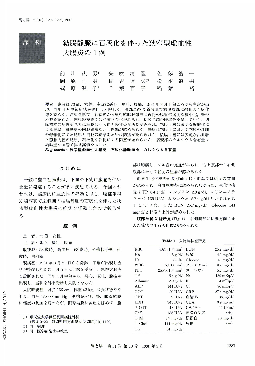
要旨 患者は73歳,女性.主訴は悪心,嘔吐,腹痛.1994年3月下旬ごろから主訴が出現.同年4月中旬症状が悪化し入院した.腹部単純X線写真で右側腹部に線状の石灰化像を認めた.注腸造影で上行結腸から横行結腸脾彎曲部近傍の腸管の著明な狭小化,壁の不整を認めた.内視鏡検査では浮腫状変化がみられ,粘膜色調が暗黒色を呈していた.切除標本の病理所見では粘膜はうっ血と慢性炎症所見がみられ,粘膜下層は著明な線維化による肥厚,細動脈の内腔狭窄ないし閉塞が認められた.動脈は粘膜下において内膜の浮腫や線維化による肥厚と内腔の狭窄あるいは閉塞が認められた.漿膜下層には広範な出血層と静脈内腔の肥厚,石灰化や骨化による閉塞が認められた.病変部のカルシウム含有量は結腸壁や血管で異常高値を示した.
A 73-year-old female was admitted to our institute with nausea, vomiting and abdominal pain which had lasted for over one month, in the middle of April, 1994. An abdominal X-ray examination revealed some lineal calcifications in the right flank. Barium enema with double contrast method showed narrowed and serrated segments from the ascending colon to the splenic flexor. A colonoscopic examination revealed edematous and melanotic coli like colored mucosa at the segments, but no malignant tissue was seen in the biopsy specimens which were obtained. Histopathological examinations of the surgically resected specimen disclosed capillary congestion and chronic inflammatory changes in the mucosa. The submucosal vessels showed severe stenosis or obstruction. The mesenteric vessels showed also arteriosclerotic stenosis and phlebosclerotic obstruction and some of them showed calcification, ossification and recanalization. The degree of calcium deposition in the resected colon tissue as well as in the mesenteric vessels was remarkably high.

Copyright © 1996, Igaku-Shoin Ltd. All rights reserved.


