Japanese
English
- 有料閲覧
- Abstract 文献概要
- 1ページ目 Look Inside
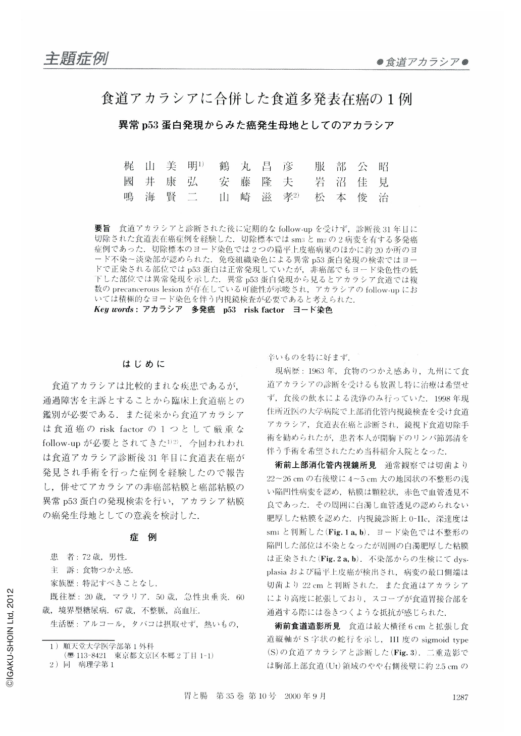
要旨 食道アカラシアと診断された後に定期的なfollow-upを受けず,診断後31年目に切除された食道表在癌症例を経験した.切除標本ではsm3とm2の2病変を有する多発癌症例であった.切除標本のヨード染色では2つの扁平上皮癌病巣のほかに約20か所のヨード不染~淡染部が認められた.免疫組織染色による異常p53蛋白発現の検索ではヨードで正染される部位ではp53蛋白は正常発現していたが,非癌部でもヨード染色性の低下した部位では異常発現を示した.異常p53蛋白発現から見るとアカラシア食道では複数のprecancerous lesionが存在している可能性が示唆され,アカラシアのfollow-upにおいては積極的なヨード染色を伴う内視鏡検査が必要であると考えられた.
We report here a case of multiple superficial cancers with achalasia, 31 years following diagnosis as achalasia. The resected specimen showed two superficial cancers with a depth of sm3 and m2. The iodine spraying method revealed approximately 20 lesions with poor staining in addition to these two cancers. From immunohistochemical staining examination with p53 monoclonal antibody, esophageal mucosa with normal staining by iodine showed normal expression of p53 protein. However, dysplastic lesions with poor staining by iodine, other than the two cancers, showed overexpression of p53 protein. We can conclude that there may be many pre-cancerous lesions in the esophagus of this achalasia patient from p53 overexpression analysis. We should follow up esophageal achalasia patients with close endoscopic examination with iodine spray.

Copyright © 2000, Igaku-Shoin Ltd. All rights reserved.


