Japanese
English
- 有料閲覧
- Abstract 文献概要
- 1ページ目 Look Inside
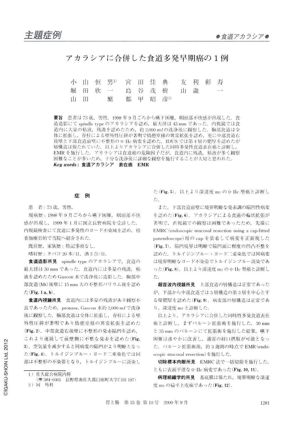
要旨 患者は73歳,男性.1998年9月ごろから嚥下困難,咽頭部不快感が出現した.食道造影にてspindle typeのアカラシアを認め,最大径は45mmであった.内視鏡では食道内に大量の粘液,残渣を認めたため,約2,000mlの洗浄後に観察した.胸部食道は全体に拡張し,脊柱による壁外性圧排が著明で偽憩室様の異常拡張を認め,更に中部食道右後壁と下部食道前壁に不整形の0-Ⅱc病変を認めた.IDUSでは第4層の肥厚を認めたが層構造は保たれていた.以上よりアカラシアに合併した同時多発性食道表在癌と診断し,EMRを施行した.アカラシアは食道癌の危険因子だが,食道内に残渣,粘液が多く観察困難なことが多いため,十分な洗浄後に詳細な観察を施行することが大切と思われた.
A 73-old-man came to our hospital, presenting esophageal dysphasia. Esophagoscopy revealed an esophageal dilatation, 45 mm in width. Esophagoscopy revealed massive amounts of mucous and residual foods in the esophageal lumen. After washing away the esophageal mucosa with about 2,000 ml water, two 0-IIc lesions were found. IDUS revealed thickness of the 4th layer without irregular layer structure, so we diagnosed the disease as esophageal multiple superficial cancer with achalasia. The dysphasia improved after balloon dilatation. Then we performed endoscopic mucosal resection for the cancer. Patients with achalasia are at a markedly increased risk of developing esophageal cancer, but, with achalasia, there are usually massive amounts of mucous and residual foods in the esophageal lumen, so careful observation should be made after washing the esophageal mucosa.
Sometimes precise observation is difficult because the esophageal lumen in achalasia tends to be winding. If observation is difficult, a fitting cap at the top of the scope will improve the accuracy.

Copyright © 2000, Igaku-Shoin Ltd. All rights reserved.


