Japanese
English
- 有料閲覧
- Abstract 文献概要
- 1ページ目 Look Inside
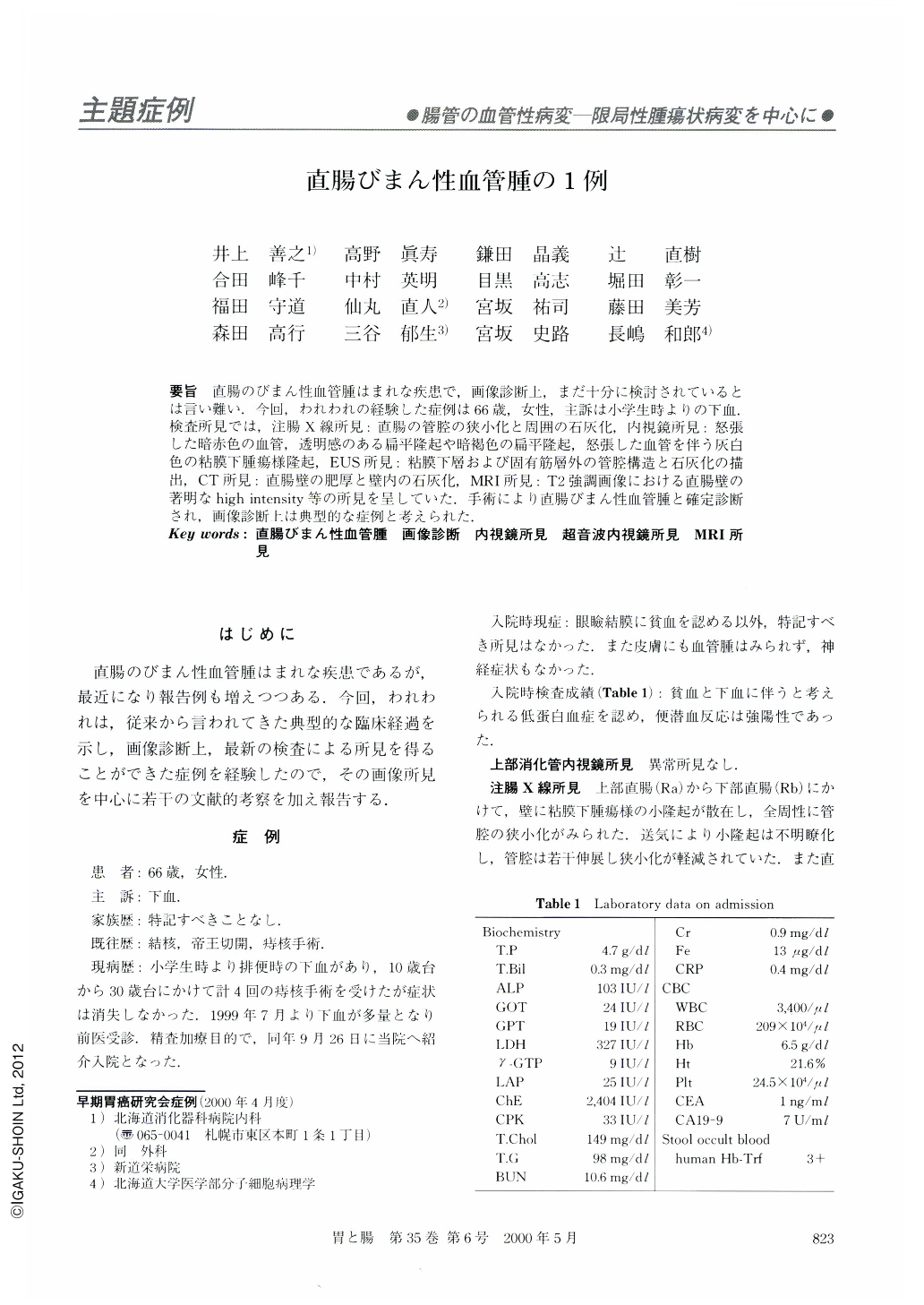
要旨 直腸のびまん性血管腫はまれな疾患で,画像診断上,まだ十分に検討されているとは言い難い.今回,われわれの経験した症例は66歳,女性,主訴は小学生時よりの下血.検査所見では,注腸X線所見:直腸の管腔の狭小化と周囲の石灰化,内視鏡所見:怒張した暗赤色の血管,透明感のある扁平隆起や暗褐色の扁平隆起,怒張した血管を伴う灰白色の粘膜下腫瘍様隆起,EUS所見:粘膜下層および固有筋層外の管腔構造と石灰化の描出,CT所見:直腸壁の肥厚と壁内の石灰化,MRI所見:T2強調画像における直腸壁の著明なhigh intensity等の所見を呈していた.手術により直腸びまん性血管腫と確定診断され,画像診断上は典型的な症例と考えられた.
A 66-year-old woman was admitted with the complaint of melena. Double contrast barium enema demonstrated diffuse narrowing of the rectal lumen with small extramural compressions. Small calcifications were seen on the rectal wall. Endoscopic findings of the rectum. showed dark-red vessels with mild dilatation, limpid flat elevation and dark-brown flat elevations. Also, many light gray “submucosal tumor” like lesions were seen with congestive vessels on their surface. Endoscopic ultrasonography (20 MHz) revealed minute duct-like structures that corresponded to the dilated vessels, and minute calcifications in the rectal wall induced a strong echo with acoustic shadowing. CT showed a thickened rectal wall with small calcifications. T2-weighted MR image of the rectal wall revealed remarkably high intensity. This patient was treated by recto-sigmoidectomy. Final histological diagnosis was hemanginoma of the rectum. In this report, we successfully demonstrated typical features of rectal hemangioma using several imaging diagnostic modalities.

Copyright © 2000, Igaku-Shoin Ltd. All rights reserved.


