Japanese
English
- 有料閲覧
- Abstract 文献概要
- 1ページ目 Look Inside
- サイト内被引用 Cited by
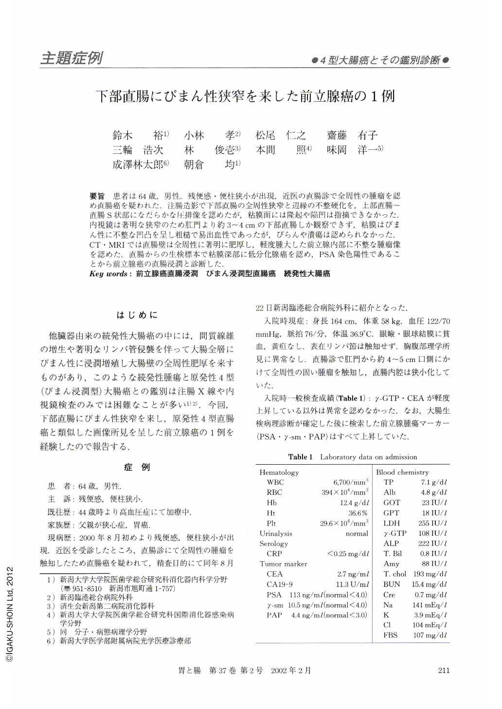
要旨 患者は64歳,男性.残便感・便柱狭小が出現,近医の直腸診で全周性の腫瘤を認め直腸癌を疑われた.注腸造影で下部直腸の全周性狭窄と辺縁の不整硬化を,上部直腸~直腸S状部になだらかな圧排像を認めたが,粘膜面には隆起や陥凹は指摘できなかった.内視鏡は著明な狭窄のため肛門より約3~4cmの下部直腸しか観察できず,粘膜はびまん性に不整な凹凸を呈し粗糙で易出血性であったが,びらんや潰瘍は認められなかった.CT・MRIでは直腸壁は全周性に著明に肥厚し,軽度腫大した前立腺内部に不整な腫瘤像を認めた.直腸からの生検標本で粘膜深部に低分化腺癌を認め,PSA染色陽性であることから前立腺癌の直腸浸潤と診断した.
A 64-year-old man suffering with residual stool and narrowing of his stools was admitted on suspicion of rectal cancer diagnosed by anal digital examination by a medical practitioner. The barium enema study showed an annular narrowing of the lower rectum with rigid contours, about 4 to 5 cm in length, and slight extrinsic compression of the upper rectum and rectosigmoid. The colonoscopic examination revealed narrow, constricted lumen of the lower rectum with edematous, nodular mucosa. There were no apparent superficial mucosal erosions or ulcers. The CT and MRI examination of the pelvis showed diffuse thickening of the rectal wall and slight enlargement of the prostate with an irregular mass in its right lobe. The specimen of the rectal biopsy showed poorly differentiated adenocarcinoma located mainly in the deeper part of the mucosa and in the submucosa. Because the tumor cells were positive for immunostaining of prostate-specific antigen, the diagnosis of prostate cancer with rectal involvement was made.

Copyright © 2002, Igaku-Shoin Ltd. All rights reserved.


