Japanese
English
- 有料閲覧
- Abstract 文献概要
- 1ページ目 Look Inside
- サイト内被引用 Cited by
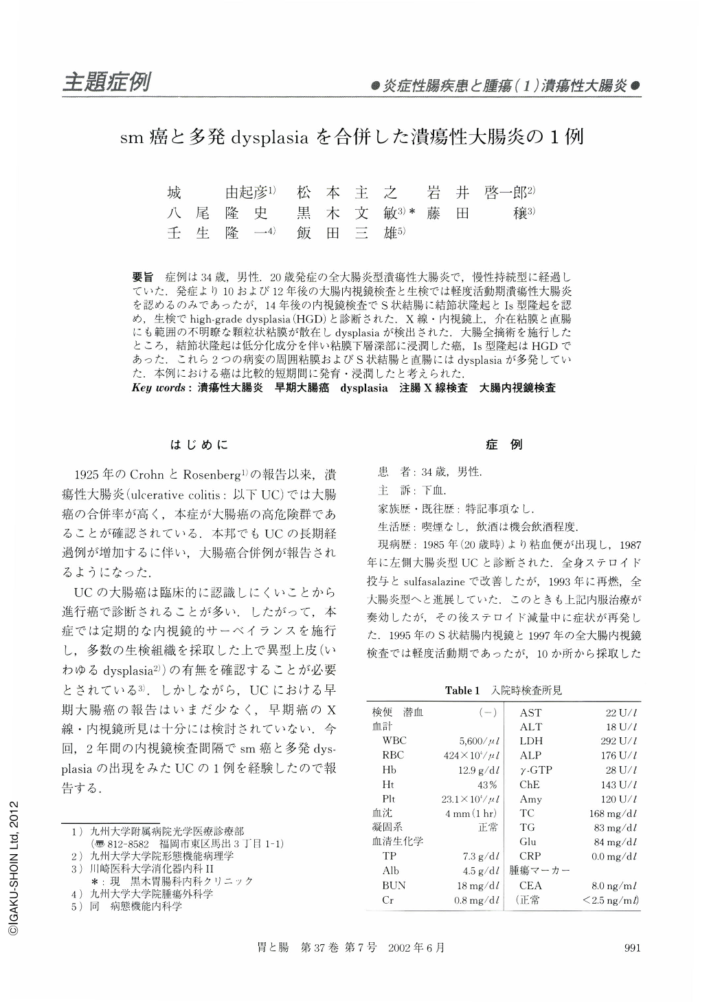
要旨 症例は34歳,男性.20歳発症の全大腸炎型潰瘍性大腸炎で,慢性持続型に経過していた.発症より10および12年後の大腸内視鏡検査と生検では軽度活動期潰瘍性大腸炎を認めるのみであったが,14年後の内視鏡検査でS状結腸に結節状隆起とⅠs型隆起を認め,生検でhigh-grade dysplasia(HGD)と診断された.X線・内視鏡上,介在粘膜と直腸にも範囲の不明瞭な顆粒状粘膜が散在しdysplasiaが検出された.大腸全摘術を施行したところ,結節状隆起は低分化成分を伴い粘膜下層深部に浸潤した癌,Ⅰs型隆起はHGDであった.これら2つの病変の周囲粘膜およびS状結腸と直腸にはdysplasiaが多発していた。本例における癌は比較的短期間に発育・浸潤したと考えられた.
A 34-year-old man with a 14-year history of ulcerative colitis was admitted in 1999, because colonoscopy revealed cancers in the sigmoid colon. Colonoscopies performed two and four years previously had not detected any dysplasia. Detailed colonoscopy and barium enema examination revealed a broad-based protrusion and a sessile polypoid lesion in the sigmoid colon, both of which were diagnosed as high-grade dysplasia. In addition, there were areas of granular mucosa where dysplasia was detected. Macroscopic examination of the resected colorectum revealed a nodular lesion and a polypoid lesion in the sigmoid colon. Histologically, the former lesion was composed of well to poorly differentiated adenocarcinoma invading the deep layer of the submucosa, and the latter lesion was high-grade dysplasia. There were also areas of low and high-grade dysplasia in the rectosigmoid colon. A comparison of histologic and colonoscopic or radiographic findings suggested that the dysplasia was depicted as granular areas. The invasive cancer in our case seems to have developed during a period of two years.

Copyright © 2002, Igaku-Shoin Ltd. All rights reserved.


