Japanese
English
- 有料閲覧
- Abstract 文献概要
- 1ページ目 Look Inside
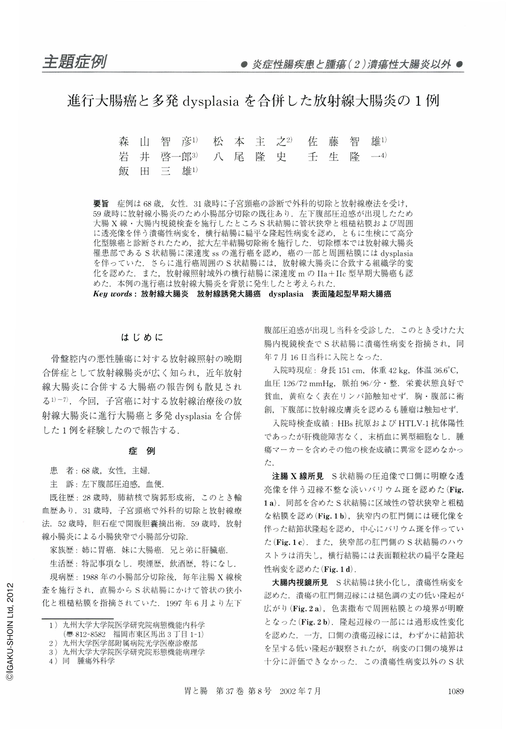
要旨 症例は68歳,女性.31歳時に子宮頸癌の診断で外科的切除と放射線療法を受け,59歳時に放射線小腸炎のため小腸部分切除の既往あり.左下腹部圧迫感が出現したため大腸X線・大腸内視鏡検査を施行したところS状結腸に管状狭窄と粗糙粘膜および周囲に透亮像を伴う潰瘍性病変を,横行結腸に扁平な隆起性病変を認め,ともに生検にて高分化型腺癌と診断されたため,拡大左半結腸切除術を施行した.切除標本では放射線大腸炎罹患部であるS状結腸に深達度ssの進行癌を認め,癌の一部と周囲粘膜にはdysplasiaを伴っていた.さらに進行癌周囲のS状結腸には,放射線大腸炎に合致する組織学的変化を認めた,また,放射線照射域外の横行結腸に深達度mのⅡa+Ⅱc型早期大腸癌も認めた.本例の進行癌は放射線大腸炎を背景に発生したと考えられた.
A 68-year-old woman with a history of irradiation for uterine cervical cancer was admitted to our institute, because of abdominal distension. Barium enema examination and total colonoscopy revealed narrowing, irregular mucosa and an ulcerating tumor in the sigmoid colon and a flat elevation in the transverse colon. Biopsy specimens from these tumors contained adenocarcinoma. Histological examination of the resected colon revealed the tumor in the sigmoid colon to be a well-differentiated adenocarcinoma invading the subserosa and that in the transverse colon to be an intramucosal adenocarcinoma. There were also areas of low or high grade dysplasia in the sigmoid colon. Histological findings compatible with radiation colitis were found in the sigmoid colon. These clinicopathologic features suggested a diagnosis of colonic cancer associated with radiation colitis.

Copyright © 2002, Igaku-Shoin Ltd. All rights reserved.


