Japanese
English
- 有料閲覧
PPV購入案内
1,320円 (1,200円+税10%) こちらは、1199ページから1207ページの文献です。購入には医書.jp本会員登録が必要です。
会員登録完了後に改めて上記「購入サイトへ」を選択してください。
または「購入サイトへ」選択後に「購入ログイン」、「新規会員登録」の順に進んでください。
会員登録について詳しくはをご確認ください。
- Abstract 文献概要
- 1ページ目 Look Inside
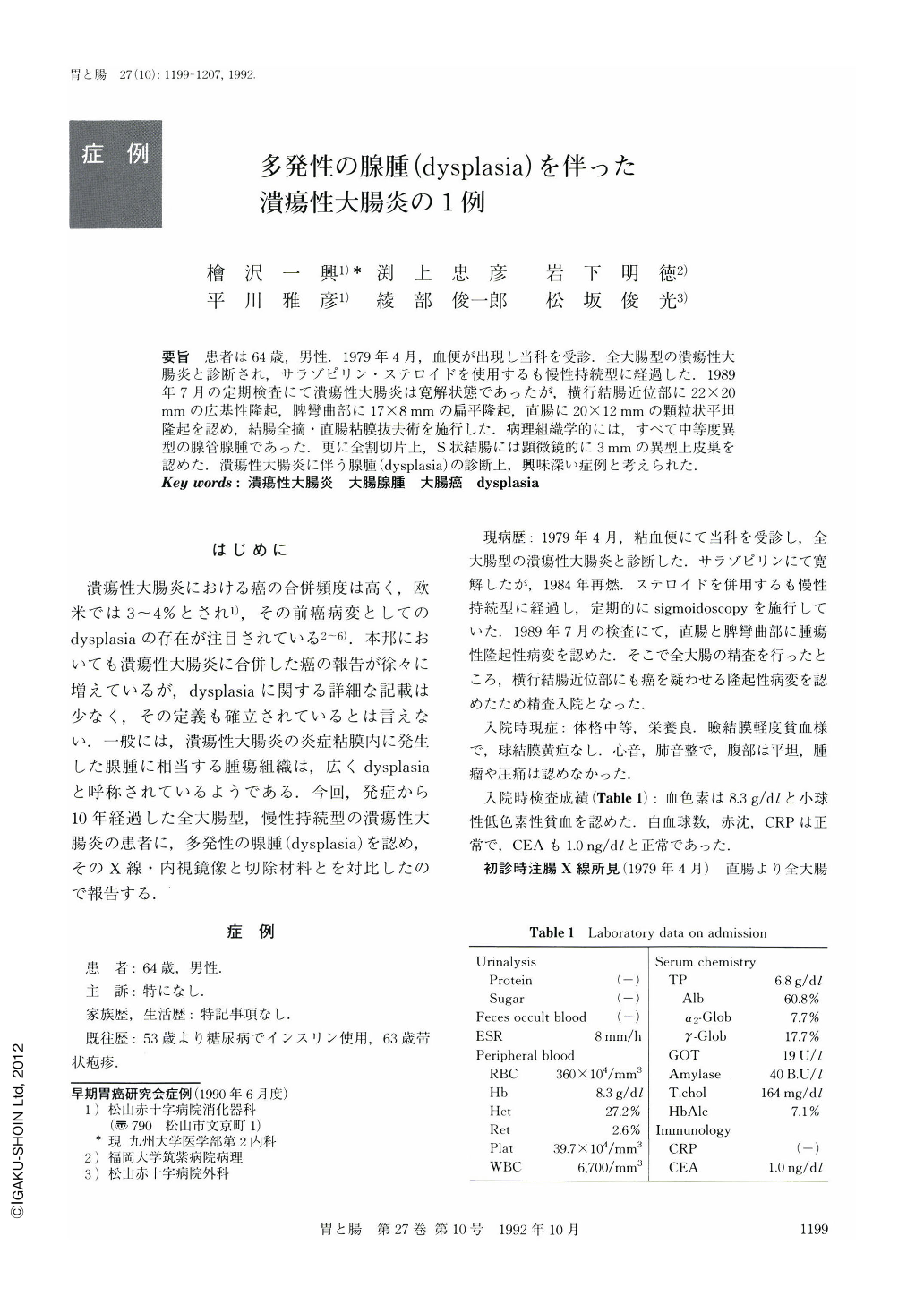
要旨 患者は64歳,男性.1979年4月,血便が出現し当科を受診.全大腸型の潰瘍性大腸炎と診断され,サラゾピリン・ステロイドを使用するも慢性持続型に経過した.1989年7月の定期検査にて潰瘍性大腸炎は寛解状態であったが,横行結腸近位部に22×20mmの広基性隆起,脾彎曲部に17×8mmの扁平隆起,直腸に20×12mmの顆粒状平坦隆起を認め,結腸全摘・直腸粘膜抜去術を施行した.病理組織学的には,すべて中等度異型の腺管腺腫であった.更に全割切片上,S状結腸には顕微鏡的に3mmの異型上皮巣を認めた.潰瘍性大腸炎に伴う腺腫(dysplasia)の診断上,興味深い症例と考えられた.
We report a case of a 64-year-old man with multifocal dysplasia of the colon after extensive ulcerative colitis of 10 years duration. He was diagnosed with ulcerative colitis (Fig. 1) in April 1979, and has repeated remissions and relapses thereafter. Barium enema (Figs. 2 and 3) and colonoscopic exmination in July 1989 revealed three elevated lesions: a slightly reddish polypoid lesion, 22×20 mm in size, in the proximal transverse colon, a plaque-like lesion, 17×8 mm in size, in the proximal descending colon, and a whitish flat granular lesion, 20×12 mm in size, in the rectum. Total proctocolectomy was performed in August 1989. Histological examination revealed these three lesions were tubular adenomas with moderate epithelial atypia, surrounded by many crypt abscesses. In addition to these lesions, an area of small flat dysplastic epithelium was detected microscopically in the sigmoid colon (Fig. 6).
Copyright © 1992, Igaku-Shoin Ltd. All rights reserved.