Japanese
English
- 有料閲覧
- Abstract 文献概要
- 1ページ目 Look Inside
- サイト内被引用 Cited by
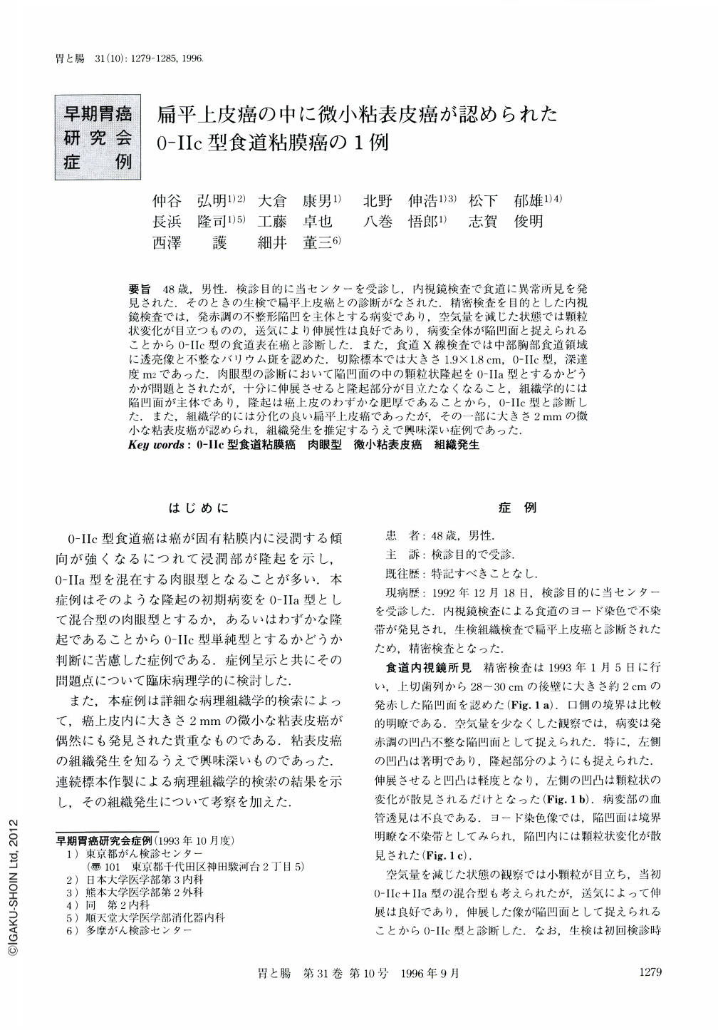
要旨 48歳,男性.検診目的に当センターを受診し,内視鏡検査で食道に異常所見を発見された.そのときの生検で扁平上皮癌との診断がなされた.精密検査を目的とした内視鏡検査では,発赤調の不整形陥凹を主体とする病変であり,空気量を減じた状態では顆粒状変化が目立つものの,送気により伸展性は良好であり,病変全体が陥凹面と捉えられることから0-Ⅱc型の食道表在癌と診断した.また,食道X線検査では中部胸部食道領域に透亮像と不整なバリウム斑を認めた.切除標本では大きさ1.9×1.8cm,0-Ⅱc型,深達度m2であった.肉眼型の診断において陥凹面の中の顆粒状隆起を0-Ⅱa型とするかどうかが問題とされたが,十分に伸展させると隆起部分が目立たなくなること,組織学的には陥凹面が主体であり,隆起は癌上皮のわずかな肥厚であることから,0-Ⅱc型と診断した.また,組織学的には分化の良い扁平上皮癌であったが,その一部に大きさ2mmの微小な粘表皮癌が認められ,組織発生を推定するうえで興味深い症例であった.
A 48-year-old male was admitted to our center for the purpose of cancer detection. On endoscopic examination of the esophagus, an iodine-unstained lesion, measuring about 2.0 cm in size, was found at the posterior of the left wall in the middle intrathoracic portion. The lesion was histologically diagnosed as Squamous cell carcinoma from the biopsy specimen.
On close endoscopic examination, when the esophageal wall was slightly extended the lesion appeared as a reddish depression with several granules, which were suspected to be 0-Ⅱc+Ⅱa type, the figure appeared to be only a depressed lesion, which was diagnosed as 0-Ⅱc type carcinoma. On close radiological examination, several granules were detected, and a small part of the lesion was depression slightly. Some clinicians diagnosed 0-Ⅱa+Ⅱc type carcinoma, but histopathologically the lesion was 0-Ⅱc type, and small granules were revealed as the thickening of cancerous epithelium and regenerative epithelium. From these results, we infer that the macroscopic figure should be diagnosed through endoscopic examination with much extension of the wall.
By detailed histopathological examination, a minute mucoepidermoid carcinoma, measuring about 2 mm in size, was found in this lesion. From the serial cuttings of the minute carcinoma, it was revealed that cancers had arisen from the lower portion of squamaous epithelium very near the esophageal duct. A small cancer nest was found 1 mm from the main nest, which caused us to suspect multiple foci carcinogenesis. It was a very valuable case for investigating the histogenesis of mucoepidermoid carcinoma.

Copyright © 1996, Igaku-Shoin Ltd. All rights reserved.


