Japanese
English
- 有料閲覧
- Abstract 文献概要
- 1ページ目 Look Inside
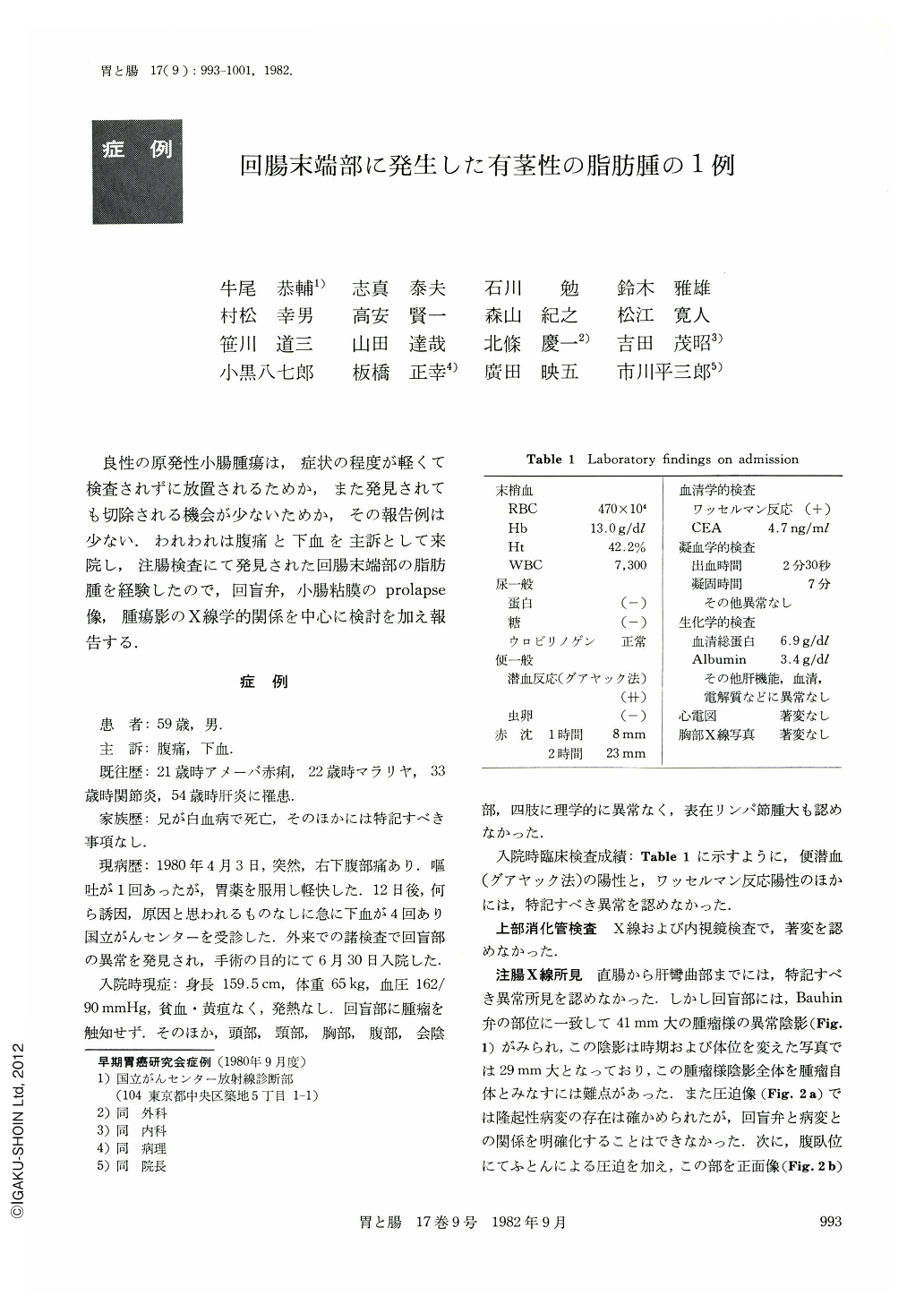
良性の原発性小腸腫瘍は,症状の程度が軽くて検査されずに放置されるためか,また発見されても切除される機会が少ないためか,その報告例は少ない.われわれは腹痛と下血を主訴として来院し,注腸検査にて発見された回腸末端部の脂肪腫を経験したので,回盲弁,小腸粘膜のprolapse像,腫瘍影のX線学的関係を中心に検討を加え報告する.
A 59-year-old man was admitted on June 30, 1980, with a history of abdominal pain, vomiting and melena. On physical examination, the abdomen was soft and there was no tenderness, or enlarged visceral palpable mass. Routine laboratory investigation did not reveal any abnormal findings except positive hemooccult reaction in feces. Upper gastrointestinal examination did not indicate any organic lesions. However, barium enema study disclosed a tumor near the ileocecal valve. Flexible fiberoptic colonoscopy revealed the tumor as a pedunculated polypoid lesion with lobulated head, hyperemic surface and central ulceration covered with yellowish white substance at the apex. The polypoid mass moved freely and was displaced behind the ileocecal valve with peristaltic wave and air pressure. Specimens taken from the surface of the tumor showed superficial erosion with inflammatory cell infiltration and fibrinous exudate. It was difficult to diagnose the tumor as a submucosal tumor before operation.
At laparotomy performed on July 9, 1980, the tumor was found in the cecum with slight ileocecal intussu-sception, which was reduced without difficulty by pres-sure, and was also detected to be pedunculated with a pedicle which originated at the wall of the terminal ileum. The tumor was removed. The cut surface was homogenous and yellowish. Pathological examination revealed a pedunculated mass which proved to be a submucosal tumor composed of mature adipose tissue with focal superficial erosion and submucosal fibrosis of surrounding mucosa.
The postoperative coures was uneventful and the patient was discharged on 29th day after operation.
Copyright © 1982, Igaku-Shoin Ltd. All rights reserved.