Japanese
English
- 有料閲覧
- Abstract 文献概要
- 1ページ目 Look Inside
- サイト内被引用 Cited by
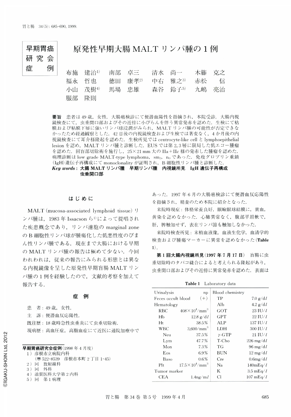
要旨 患者は49歳,女性.大腸癌検診にて便潜血陽性を指摘され,本院受診.大腸内視鏡検査にて,虫垂開口部およびその近傍に小びらんを伴う異常発赤を認めた.生検にて粘膜および粘膜下層に強いリンパ球浸潤がみられ,MALTリンパ腫の可能性が否定できなかったため経過観察とした.42日後の内視鏡検査および生検では著変なく,4か月後の内視鏡検査にて耳介様隆起を認めた。生検所見ではcentrocyte-like cellとlymphoepithelial lesionを認め,MALTリンパ腫と診断した.EUSでは第2,3層に限局した低エコー腫瘤を認めた.回盲部切除術を施行し,25×21mm大のⅡa+Ⅱc様の発赤した腫瘤を認めた.病理診断はlow grade MALT-type lymphoma,sm2,n0であった.免疫グロブリン重鎖(IgH)遺伝子再構成にてmonoclonalityが証明され,B細胞性リンパ腫と診断した.
A 49-year-old woman attended our hospital because of a positive fecal occult blood test. Colonoscopy showed an abnormal reddish lesion with small erosions near the orifice of the appendix. Mucosa-associated lymphoid tissue (MALT) lymphoma could not be ruled out, because biopsy specimens revealed marked lymphocytic infiltration in the mucosa and submucosa. After 42 days, repeat endoscopy and biopsy showed no marked changes. Approximately four months after the initial examination, the third endoscopy revealed a pinna-like elevation, and biopsy disclosed centrocytelike cells and lymphoepithelial lesions. So the lesion was diagnosed as MALT lymphoma. Endoscopic ultrasonography revealed a hypoechoic mass in the second and third layers of the bowel wall. Ileocecal resection was performed, and the resected specimen contained a rid type Ⅱa + Ⅱc-like lesion measuring 25×21 mm. Histological examination revealed low-grade MALT lymphoma (sm2, n0). A diagnosis of B-cell lymphoma was made by detecting monoclonal rearrangement of the immunoglobulin heavy chain gene.
Copyright © 1999, Igaku-Shoin Ltd. All rights reserved.