Japanese
English
- 有料閲覧
- Abstract 文献概要
- 1ページ目 Look Inside
- サイト内被引用 Cited by
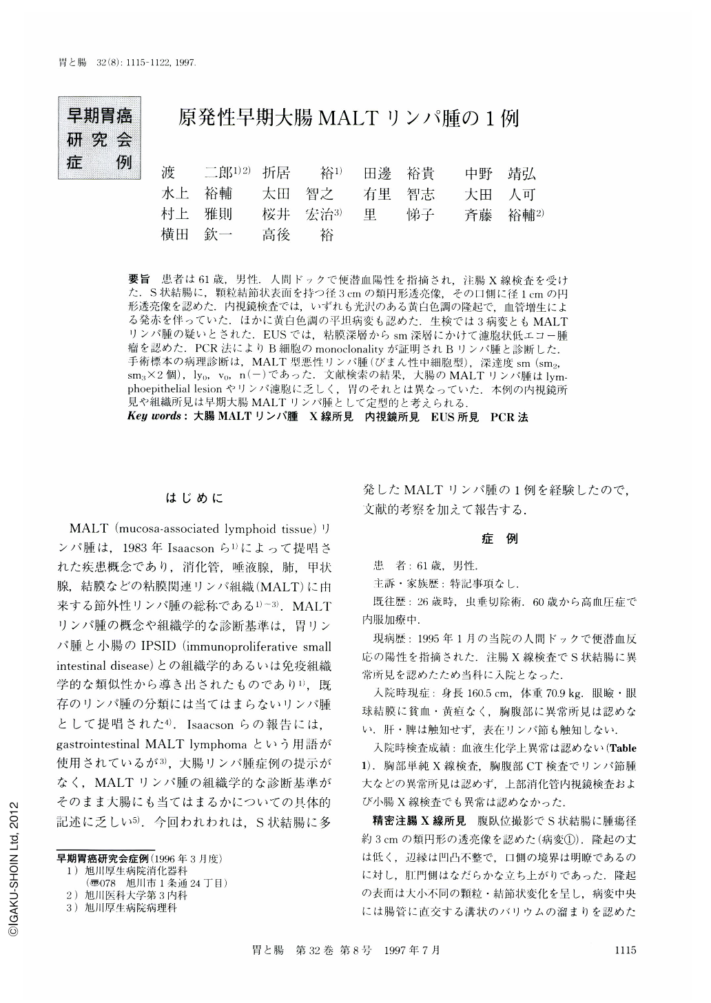
要旨 患者は61歳,男性.人間ドックで便潜血陽性を指摘され,注腸X線検査を受けた.S状結腸に,顆粒結節状表面を持つ径3cmの類円形透亮像,その口側に径1cmの円形透亮像を認めた.内視鏡検査では,いずれも光沢のある黄白色調の隆起で,血管増生による発赤を伴っていた.ほかに黄白色調の平坦病変も認めた.生検では3病変ともMALTリンパ腫の疑いとされた.EUSでは,粘膜深層からsm深層にかけて濾胞状低エコー腫瘤を認めた,PCR法によりB細胞のmonoclonalityが証明されBリンパ腫と診断した.手術標本の病理診断は,MALT型悪性リンパ腫(びまん性中細胞型),深達度sm(sm2,sm3×2個),ly0,v0,n(-)であった.文献検索の結果,大腸のMALTリンパ腫はlymphoepithelial lesionやリンパ濾胞に乏しく,胃のそれとは異なっていた.本例の内視鏡所見や組織所見は早期大腸MALTリンパ腫として定型的と考えられる.
A 61-year-old man received a close examination of the colon because feces' occult blood test was positive during a physical check-up in our hospital. Barium enema study showed two polypoid lesions in the sigmoid colon, one of which was a flat elevation measuring 30 mm in diameter with a granular to nodular surface, the other was a hemispherical polyp measuring 10 mm in diameter. In colonoscopic observation, these tumors showed a glossy yellowish-white surface with capillary proliferation. A flat yellowish-white lesion was also detected in the sigmoid colon in addition to these tumors. By histologic examination of biopsy specimens, all of them were suspected of MALT (mucosa-associated lymphoid tissue) lymphoma. High-frequency ultrasound probe showed a massive invasion in the submucosal layer. A definite diagnosis of B-cell lymphoma was obtained by the polymerase chain reaction, which disclosed a monoclonal rearrangement of immunoglobulin heavy chain gene. Final diagnosis after sigmoidectomy was diffuse lymphoma, medium cell type (MALT-type). Tumor invasion was limited to the submucosa without nodal involvement. In the literature, lymphoepithelial lesions and lymphoid follicles are shown to be less prominent in MALT lymphomas of the colon than those of the stomach. Endoscopic and histologic findings in the present case seem to be typical of MALT lymphoma of the colon in the early stage, although this is extremely rare.

Copyright © 1997, Igaku-Shoin Ltd. All rights reserved.


