Japanese
English
- 有料閲覧
- Abstract 文献概要
- 1ページ目 Look Inside
- 参考文献 Reference
- サイト内被引用 Cited by
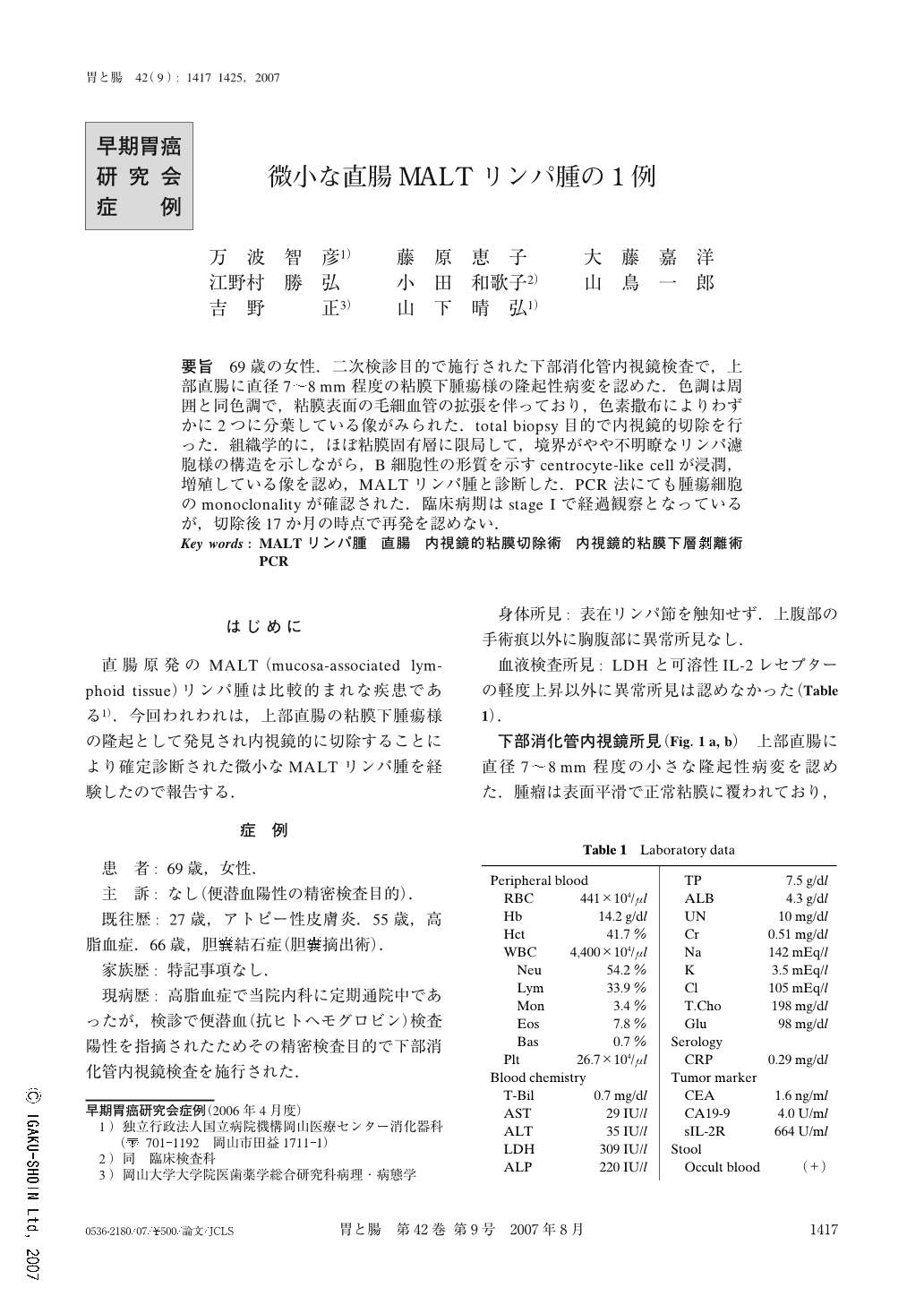
要旨 69歳の女性.二次検診目的で施行された下部消化管内視鏡検査で,上部直腸に直径7~8mm程度の粘膜下腫瘍様の隆起性病変を認めた.色調は周囲と同色調で,粘膜表面の毛細血管の拡張を伴っており,色素撒布によりわずかに2つに分葉している像がみられた.total biopsy目的で内視鏡的切除を行った.組織学的に,ほぼ粘膜固有層に限局して,境界がやや不明瞭なリンパ濾胞様の構造を示しながら,B細胞性の形質を示すcentrocyte-like cellが浸潤,増殖している像を認め,MALTリンパ腫と診断した.PCR法にても腫瘍細胞のmonoclonalityが確認された.臨床病期はstage Iで経過観察となっているが,切除後17か月の時点で再発を認めない.
A 69-year-old Japanese female underwent colonoscopy for further examination of occult blood in the stool. A sessile polypoid lesion resembling a submucosal tumor 7~8mm in diameter was found in the upper rectum. The elevation, appearing to be the same color as adjacent normal mucosa, had a smooth and even surface with telangiectasia. En bloc, complete resection was performed endoscopically. Histology revealed that the tumor was predominantly confined to the lamina propria mucosae and consisted of centrocyte-like cells that were shown by immunohistochemistry to be positive for B-cell markers. These findings were morphologically consistent with extranodal marginal zone B-cell lymphoma of mucosa-associated lymphoid tissue type (MALT lymphoma). Polymerase chain reaction (PCR) demonstrated monoclonality of the lymphoma cells. No other site of involvement was identified by the staging work-up. There is no endoscopical or histological evidence of lymphoma in the rectum after 17 months of follow-up. A minute primary MALT lymphoma of the rectum has been rarely reported.

Copyright © 2007, Igaku-Shoin Ltd. All rights reserved.


