Japanese
English
- 有料閲覧
PPV購入案内
1,320円 (1,200円+税10%) こちらは、372ページから377ページの文献です。購入には医書.jp本会員登録が必要です。
会員登録完了後に改めて上記「購入サイトへ」を選択してください。
または「購入サイトへ」選択後に「購入ログイン」、「新規会員登録」の順に進んでください。
会員登録について詳しくはをご確認ください。
- Abstract 文献概要
- 1ページ目 Look Inside
- 参考文献 Reference
- サイト内被引用 Cited by
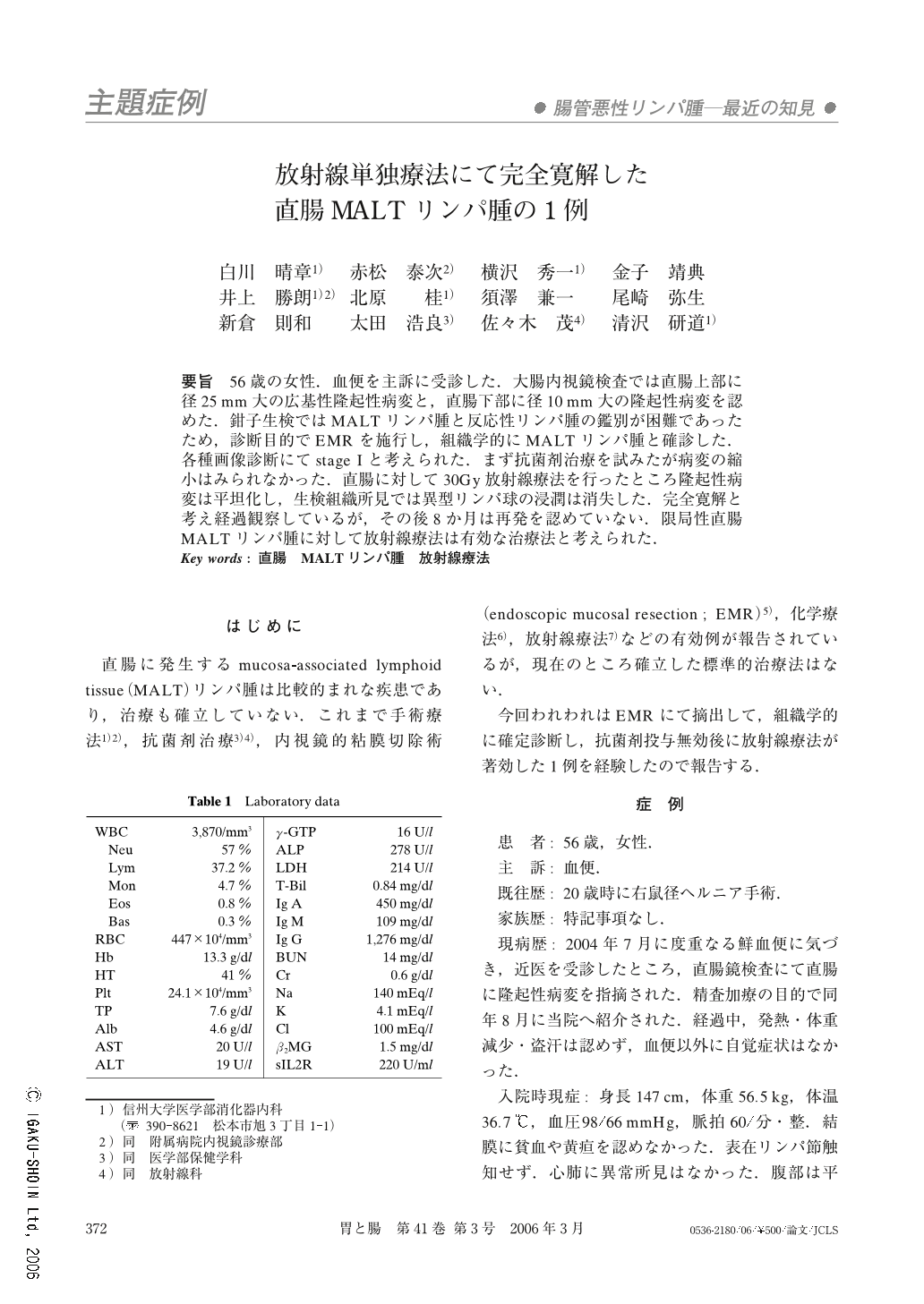
要旨 56歳の女性.血便を主訴に受診した.大腸内視鏡検査では直腸上部に径25mm大の広基性隆起性病変と,直腸下部に径10mm大の隆起性病変を認めた.鉗子生検ではMALTリンパ腫と反応性リンパ腫の鑑別が困難であったため,診断目的でEMRを施行し,組織学的にMALTリンパ腫と確診した.各種画像診断にてstage Iと考えられた.まず抗菌剤治療を試みたが病変の縮小はみられなかった.直腸に対して30Gy放射線療法を行ったところ隆起性病変は平坦化し,生検組織所見では異型リンパ球の浸潤は消失した.完全寛解と考え経過観察しているが,その後8か月は再発を認めていない.限局性直腸MALTリンパ腫に対して放射線療法は有効な治療法と考えられた.
A 56-year-old woman was referred to our hospital in August, 2004 for further examination of rectal tumors. The first colonoscopy revealed two elevated lesions ; one was a sessile elevated lesion (25mm in size) with nodular surface in the upper rectum, and the other was a small elevated lesion (10mm in size) in the lower rectum. Endoscopic ultrasonography showed thick hypoechoic change within the second layer. Histological finding of conventional biopsy could not definitively distinguish whether it was MALT lymphoma or reactive lymphoid hyperplasia. Jumbo biopsy using endoscopic mucosal resection (2 channel method) was performed for the sessile elevated lesion, and histological findings of it revealed infiltration of atypical lymphoid cells such as centrocyte-like cells in the mucosa and the submucosa. Immunohistochemical study showed that atypical lymphocytes were positive for CD20, and negative for CD3, CD5, and cyclin D1. The monoclonality of B-cells was not detected by polymerase chain reaction products for immunoglobulin heavy chain. Bone marrow examination and some imaging procedures revealed no other infiltration of lymphoma cells except at the rectum. From these findings, this patient was diagnosed as stage I rectal MALT lymphoma. Helicobacter pylori (H. pylori) was not detected in histology or culture of the patients gastric biopsy specimens. After obtaining adequately informed consent, antibiotic therapy using Levofloxacin (3×100mg/day) was administered for 7 days. However, no remarkable regression was recognized in the endoscopic and histological findings. After that, she was administered 30 Gy radiation therapy for the rectal lesion. No remarkable adverse effect except slight leukopenia was recognized. Colonoscopy after radiation therapy revealed remarkable regression of the rectal MALT lymphoma, and typical lymphoma cells had disappeared in the biopsy specimens. She has had no recurrence in the 12 months since radiation therapy.
Radiation therapy is thought to be a useful therapeutic procedure for localized rectal MALT lymphoma as it is also for localized gastric MALT lymphoma without H. pylori infection or with resistance against eradication therapy for H. Pylori.
Copyright © 2006, Igaku-Shoin Ltd. All rights reserved.