Japanese
English
- 有料閲覧
- Abstract 文献概要
- 1ページ目 Look Inside
- 参考文献 Reference
- サイト内被引用 Cited by
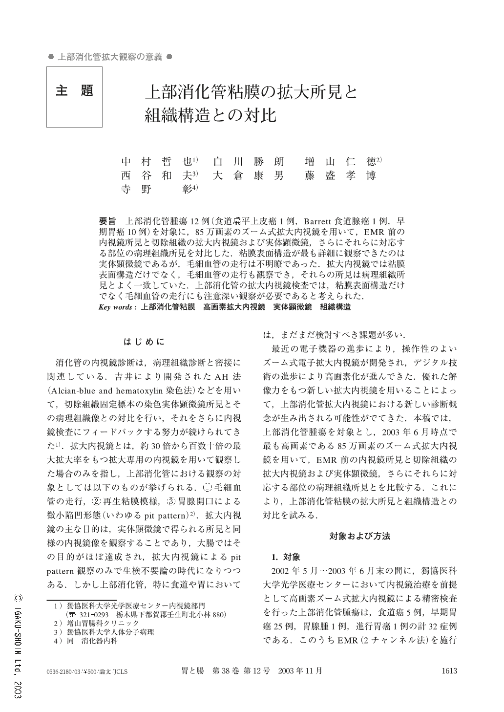
要旨 上部消化管腫瘍12例(食道扁平上皮癌1例,Barrett食道腺癌1例,早期胃癌10例)を対象に,85万画素のズーム式拡大内視鏡を用いて,EMR前の内視鏡所見と切除組織の拡大内視鏡および実体顕微鏡,さらにそれらに対応する部位の病理組織所見を対比した.粘膜表面構造が最も詳細に観察できたのは実体顕微鏡であるが,毛細血管の走行は不明瞭であった.拡大内視鏡では粘膜表面構造だけでなく,毛細血管の走行も観察でき,それらの所見は病理組織所見とよく一致していた.上部消化管の拡大内視鏡検査では,粘膜表面構造だけでなく毛細血管の走行にも注意深い観察が必要であると考えられた.
We compare the views of high-resolution magnifying endoscopy and stereoscopic microscopy with the histological findings of upper gastrointestinal tumors which were resected by endoscopic mucosal resection. In all the tumors which were examined (total 12 cases, including 10 cases of early gastric cancer, 1 case of esophageal squamous cell carcinoma, 1 case of Barrett's adenocarcinoma), we were able to observe the features of the capillary vessels of the tumors by high-resolution magnifying endoscopy. In 11 of these 12 cases (excluding the case of esophageal squamous cell carcinoma), we could also clearly observe the fine mucosal patterns of the tumors by stereoscopic microscopy, but the features of the capillary vessels were obscured. The fine mucosal patterns and the features of the capillary vessels which were observed by high-resolution magnifying endoscopy were consistent with the histological findings. High-resolution magnifying endoscopy may be very useful in predicting the histological diagnosis of upper gastrointestinal tumors.

Copyright © 2003, Igaku-Shoin Ltd. All rights reserved.


