Japanese
English
- 有料閲覧
- Abstract 文献概要
- 1ページ目 Look Inside
- 参考文献 Reference
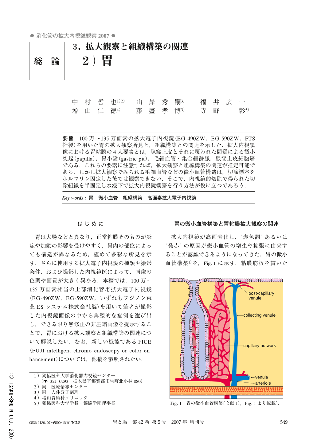
要旨 100万~135万画素の拡大電子内視鏡(EG-490ZW,EG-590ZW,FTS社製)を用いた胃の拡大観察所見と,組織構築との関連を示した.拡大内視鏡像における胃粘膜の4大要素とは,腺窩上皮とそれに覆われた間質による微小突起(papilla),胃小窩(gastric pit),毛細血管・集合細静脈,腺窩上皮細胞層である.これらの要素に注意すれば,拡大観察と組織構築の関連が推定可能である.しかし拡大観察でみられる毛細血管などの微小血管構造は,切除標本をホルマリン固定した後では観察できない.そこで,内視鏡的切除で得られた切除組織を半固定し水浸下で拡大内視鏡観察を行う方法が役に立つであろう.
We compared high-resolution (100 to 135 megapixels) magnifying endoscopic findings of the stomach with their histological structures. Histological structures such as gastric pit, capillaries, the layer of the foveolar epithelium, and the minute projections which comprise the foveolar epithelium and the lamina propria are important when we diagnose gastric lesions using magnifying endoscopy. Concerning the relations between magnifying endoscopic findings and capillaries of the resected specimen, it is useful to observe the specimen which is half fixed by formalin.

Copyright © 2007, Igaku-Shoin Ltd. All rights reserved.


