Japanese
English
- 有料閲覧
- Abstract 文献概要
- 1ページ目 Look Inside
- 参考文献 Reference
- サイト内被引用 Cited by
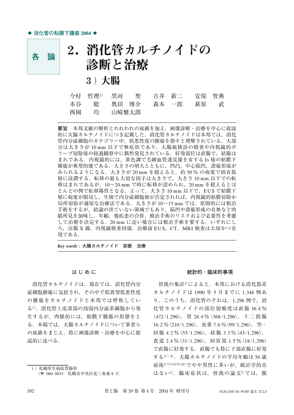
要旨 本邦文献の解析とわれわれの成績を加え,画像診断・治療を中心に総説的に大腸カルチノイドにつき記載した.消化管カルチノイドは本邦では,消化管内分泌細胞のカテゴリー中,低悪性度の腫瘍を指すと理解されている.大部分は大きさが10mm以下で無症状であり,大腸癌検診の精査や内視鏡的ポリープ切除後の経過観察中に偶然発見されている.好発部位は直腸で,結腸はまれである.内視鏡的には,黄色調で毛細血管透見像を有するIs様の粘膜下腫瘍が典型的像である.大きさの増大とともに,凹凸,中心陥凹,潰瘍形成がみられるようになる.大きさが20mmを超えると,約50%の病変で固有筋層に浸潤する.転移の最も大切な因子は大きさで,大きさ10mm以下での転移はまれであるが,10~20mmで時に転移が認められ,20mmを超えるとほとんどの例で転移陽性となる.よって,大きさ10mm以下で,EUSで粘膜下層に病変が限局し,生検で内分泌細胞癌が否定されれば,内視鏡的粘膜切除か局所切除が適切な治療法である.大きさが10~15mmでは,原則的には根治手術をするが,結論の出ていない領域でもあり,陥凹や潰瘍形成の有無など肉眼所見を加味し,年齢,他疾患の合併,根治手術のリスクおよび必要性を考慮して治療を決定する.20mmに近い場合には根治手術を要する.いずれにしろ,注腸X線,内視鏡検査同様,治療前EUS,CT,MRI検査は大切かつ有用である.
We described carcinoid tumor of the colon and rectum based on analysis of Japanese literature including our own results.
In Japan, carcinoids of the gastro-intesinal tract are thought to be in the category of endocrine cell tumors with low-grade malignancy. They are mostly asymptomatic because their size is monthly less than10mm in diameter, so most of such tumors are incidentally found in the course of colon cancer screening or in follow-up colonoscopy after endoscopic treatment. The rectum is the most common site, however, colon carcinomas are rare in our country.
Typical endoscopic view is an Is-like submucosal tumor with yellowish color and visible vascular pattern. They are apt to have uneven, central depression or ulceration on the surface and to be enlarged in size.
Over about50percent of the carcinoids beyond20mm in diameter invade is the proper muscle layer. The most important factor indicating possible metastasis is size : less than10mm is very rare, between10and20mm occasionally, greater than20mm almost always.
Therefore, as for management, if the size is under10mm, and if it is shown to be limited to the submucosa by endoscopic ultra-sonography (EUS) and not to be an endocrine cell carcinoma by biopsy, endoscopic mucosal resection or local excision is appropriate. If from10to20mm, the patient should on principle have radical operation, though, especially in the range of from10to15mm is the controversial zone for clinical treatment, and leaves room for less radical treatment based on age, complication of other diseases, operative risk and necessity of permanent colostomy, including evaluations of macroscopic findings with or without central depression or ulceration. If the size is close to20mm, radical operation is needed. EUS, CT, MRI are important and helpful as is also barium enema and colonoscopy before any management procedures are decided.
1) Division of Gastroenterology, Sapporo Kosei General Hospital, Sapporo, Japan

Copyright © 2004, Igaku-Shoin Ltd. All rights reserved.


