Japanese
English
- 有料閲覧
- Abstract 文献概要
- 1ページ目 Look Inside
- 参考文献 Reference
- サイト内被引用 Cited by
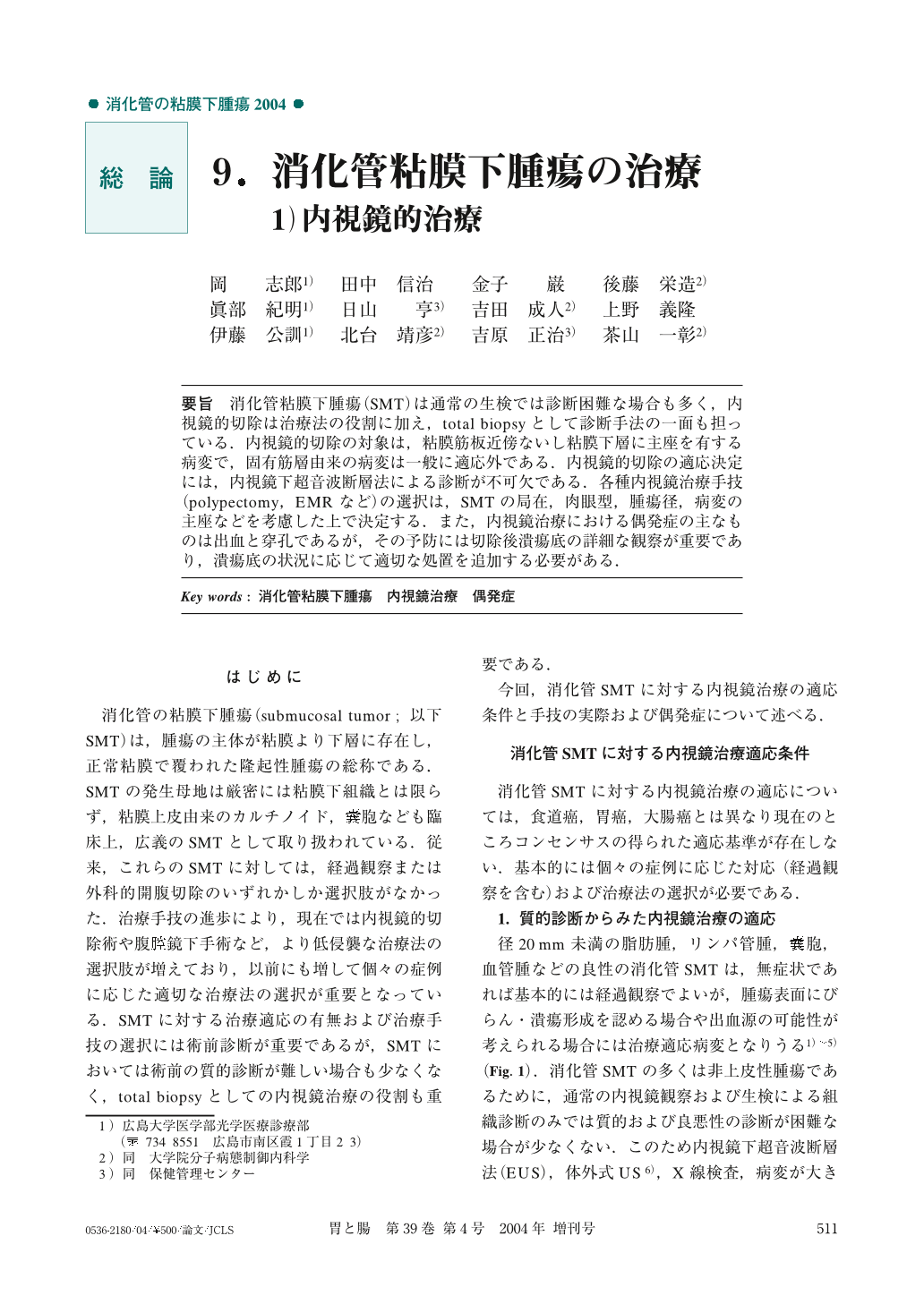
要旨 消化管粘膜下腫瘍(SMT)は通常の生検では診断困難な場合も多く,内視鏡的切除は治療法の役割に加え,total biopsyとして診断手法の一面も担っている.内視鏡的切除の対象は,粘膜筋板近傍ないし粘膜下層に主座を有する病変で,固有筋層由来の病変は一般に適応外である.内視鏡的切除の適応決定には,内視鏡下超音波断層法による診断が不可欠である.各種内視鏡治療手技(polypectomy,EMRなど)の選択は,SMTの局在,肉眼型,腫瘍径,病変の主座などを考慮した上で決定する.また,内視鏡治療における偶発症の主なものは出血と穿孔であるが,その予防には切除後潰瘍底の詳細な観察が重要であり,潰瘍底の状況に応じて適切な処置を追加する必要がある.
In many cases, it is difficult to diagnose submucosal tumor (SMT) of the GI tract by ordinary biopsy, but, added to its role as a means of treatment, endoscopic resection, by using total biopsy, endoscopic resection can also be used as a diagnostic technique. Subjects of endoscopic resection include lesions which have the main part near the lamina muscularis mucosa or in the submucosal layer, but, generally, lesions derived from the muscularis propria are not suitable indication. Diagnosis by endoscopic ultrasonic tomography is essential to determine the indication for endoscopic resection. Selection of various endoscopic treatment techniques is determined, taking into consideration localization of SMT, macroscopic type, tumor diameter, and the location of the main part of the lesions. Major accident in endoscopic treatment includes bleeding and perforation, so, in order to prevent such accidents, it is very important to observe in detail the ulcer floor after resection and it is necessary to use additional appropriate treatment according to the condition of the ulcer floor.
1) Department of Endoscopy, Hiroshima University Hospital, Hiroshima, Japan

Copyright © 2004, Igaku-Shoin Ltd. All rights reserved.


