Japanese
English
- 有料閲覧
PPV購入案内
2,750円 (2,500円+税10%) こちらは、116ページから122ページの文献です。購入には医書.jp本会員登録が必要です。
会員登録完了後に改めて上記「購入サイトへ」を選択してください。
または「購入サイトへ」選択後に「購入ログイン」、「新規会員登録」の順に進んでください。
会員登録について詳しくはをご確認ください。
- Abstract 文献概要
- 1ページ目 Look Inside
- 参考文献 Reference
要旨
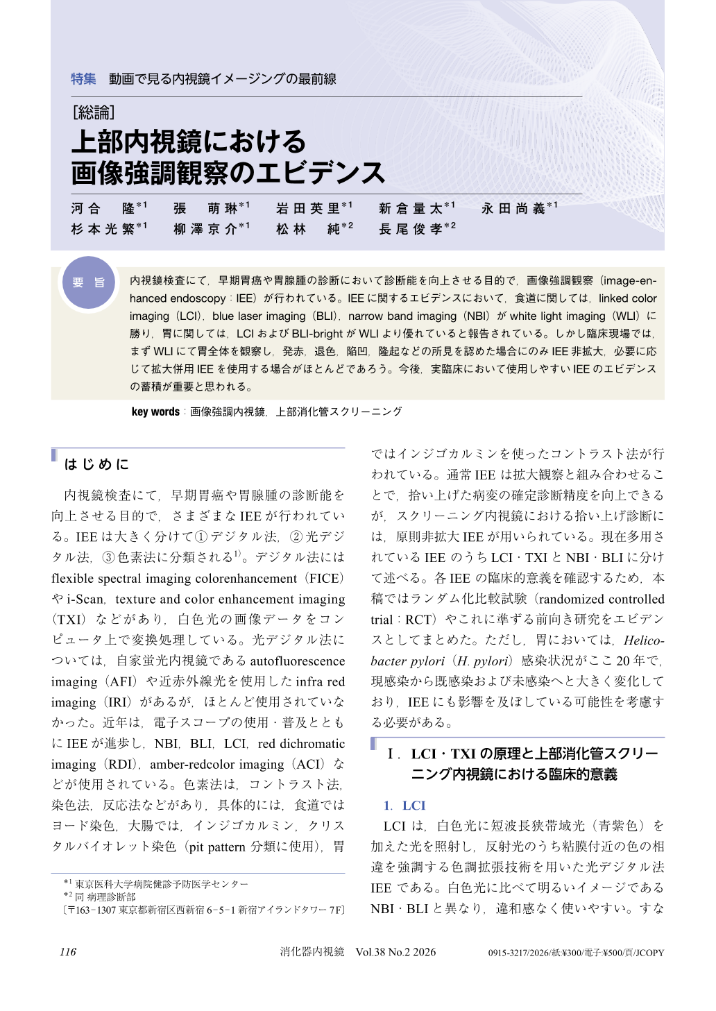
内視鏡検査にて,早期胃癌や胃腺腫の診断において診断能を向上させる目的で,画像強調観察(image-enhanced endoscopy:IEE)が行われている。IEEに関するエビデンスにおいて,食道に関しては,linked color imaging(LCI),blue laser imaging(BLI),narrow band imaging(NBI)がwhite light imaging(WLI)に勝り,胃に関しては,LCIおよびBLI-brightがWLIより優れていると報告されている。しかし臨床現場では,まずWLIにて胃全体を観察し,発赤,退色,陥凹,隆起などの所見を認めた場合にのみIEE非拡大,必要に応じて拡大併用IEEを使用する場合がほとんどであろう。今後,実臨床において使用しやすいIEEのエビデンスの蓄積が重要と思われる。
Image-enhanced endoscopy (IEE) is performed to improve diagnostic accuracy for early gastric cancer and gastric adenomas during endoscopic examinations. Evidence regarding IEE indicates that for the esophagus, linked color imaging (LCI), blue laser imaging (BLI), and narrow band imaging (NBI) outperform white light imaging (WLI). For the stomach, LCI and BLI-bright are reported to be superior to WLI. However, in clinical practice, WLI is typically used first to observe the entire stomach. IEE without magnification is used only when findings such as erythema, pale color lesions, depressive lesions, or raised lesions are detected, with magnification-enhanced IEE used as needed. Accumulating evidence for AI-based IEE that is easy to use in actual clinical practice is considered important going forward.
© tokyo-igakusha.co.jp. All right reserved.