Japanese
English
- 有料閲覧
PPV購入案内
1,650円 (1,500円+税10%) こちらは、593ページから597ページの文献です。購入には医書.jp本会員登録が必要です。
会員登録完了後に改めて上記「購入サイトへ」を選択してください。
または「購入サイトへ」選択後に「購入ログイン」、「新規会員登録」の順に進んでください。
会員登録について詳しくはをご確認ください。
- Abstract 文献概要
- 1ページ目 Look Inside
- 参考文献 Reference
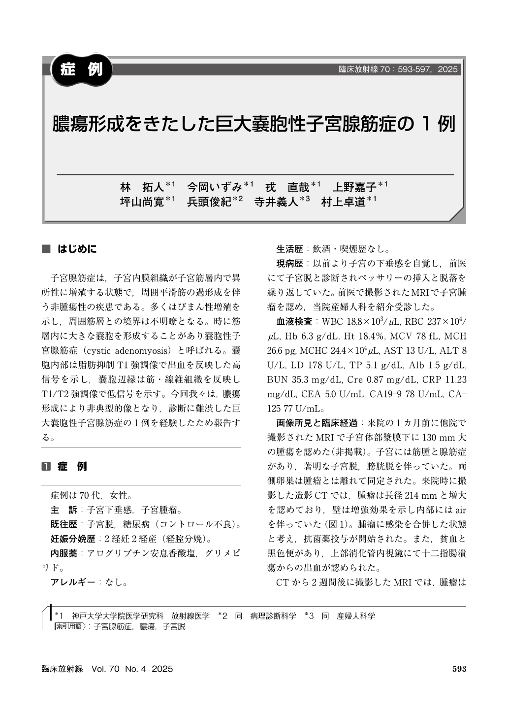
子宮腺筋症は,子宮内膜組織が子宮筋層内で異所性に増殖する状態で,周囲平滑筋の過形成を伴う非腫瘍性の疾患である。多くはびまん性増殖を示し,周囲筋層との境界は不明瞭となる。時に筋層内に大きな嚢胞を形成することがあり嚢胞性子宮腺筋症(cystic adenomyosis)と呼ばれる。嚢胞内部は脂肪抑制T1強調像で出血を反映した高信号を示し,嚢胞辺縁は筋・線維組織を反映しT1/T2強調像で低信号を示す。今回我々は,膿瘍形成により非典型的像となり,診断に難渋した巨大嚢胞性子宮腺筋症の1例を経験したため報告する。
We report a rare case of huge cystic adenomyosis with abscess. A woman in her 70s presented a rapidly growing uterine mass. She also suffered from recurrent uterine prolapse and diabetes. CT and MR imaging showed the mass was consisted of enhanced wall and large cystic space with gas, and diffusion weighed imaging suggested abscess formation. Surgery showed extensive necrosis and yellowish-green pus within the mass, though no bacterial or fungal pathogens were identified. On pathological examination, smooth muscle tissue and dilated gland-like structures were seen in the thickened wall, therefore the diagnosis of cystic adenomyosis was made. Reports of adenomyosis with abscess are rare, mechanisms nor causative organisms were often unidentified. In our case, recurrent uterine prolapse might induce ischemic to necrotic change, however, preoperative diagnosis remained difficult.
Copyright © 2025, KANEHARA SHUPPAN Co.LTD. All rights reserved.