Japanese
English
- 有料閲覧
PPV購入案内
1,650円 (1,500円+税10%) こちらは、27ページから37ページの文献です。購入には医書.jp本会員登録が必要です。
会員登録完了後に改めて上記「購入サイトへ」を選択してください。
または「購入サイトへ」選択後に「購入ログイン」、「新規会員登録」の順に進んでください。
会員登録について詳しくはをご確認ください。
- Abstract 文献概要
- 1ページ目 Look Inside
- 参考文献 Reference
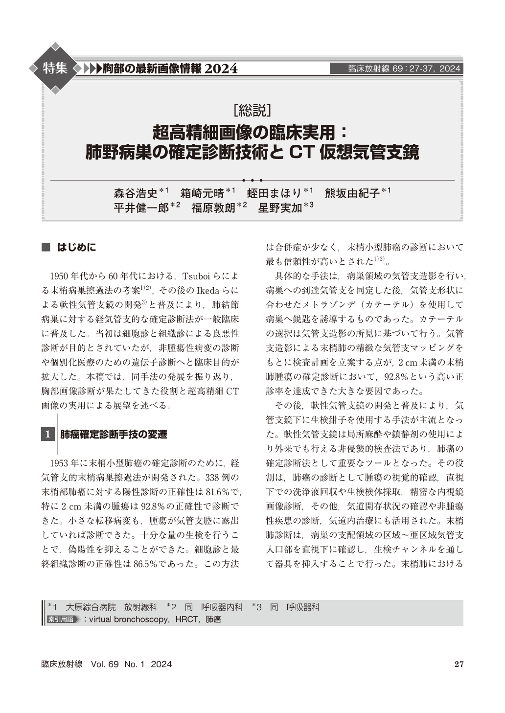
1950年代から60年代における,Tsuboiらによる末梢病巣擦過法の考案1)2),その後のIkedaらによる軟性気管支鏡の開発3)と普及により,肺結節病巣に対する経気管支的な確定診断法が一般臨床に普及した。当初は細胞診と組織診による良悪性診断が目的とされていたが,非腫瘍性病変の診断や個別化医療のための遺伝子診断へと臨床目的が拡大した。本稿では,同手法の発展を振り返り,胸部画像診断が果たしてきた役割と超高精細CT画像の実用による展望を述べる。
In the 1950s and 1960s, the development of the peripheral lesion abrasion method by Eitaka Tsuboi et al and the subsequent development and popularization of the flexible bronchoscope by Shigeto Ikeda et al.
Initially, the purpose was to diagnose benign or malignant lesions using cytology and histology, but now the purpose has expanded to include diagnosis of nonneoplastic lesions and genetic diagnosis for personalized medicine.
Prior information on bronchial bifurcation morphology is useful for transbronchial device approaches to lung lesions. In recent years, high-resolution CT has made it possible to perform precise virtual bronchoscopy, which has improved the accuracy of definitive diagnosis of lung lesions.
This paper reviews the development of the method, and discusses the role that thoracic imaging has played and the prospects with the practical application of ultrahigh-resolution CT images.
Copyright © 2024, KANEHARA SHUPPAN Co.LTD. All rights reserved.