Japanese
English
- 有料閲覧
- Abstract 文献概要
- 1ページ目 Look Inside
- 参考文献 Reference
はじめに
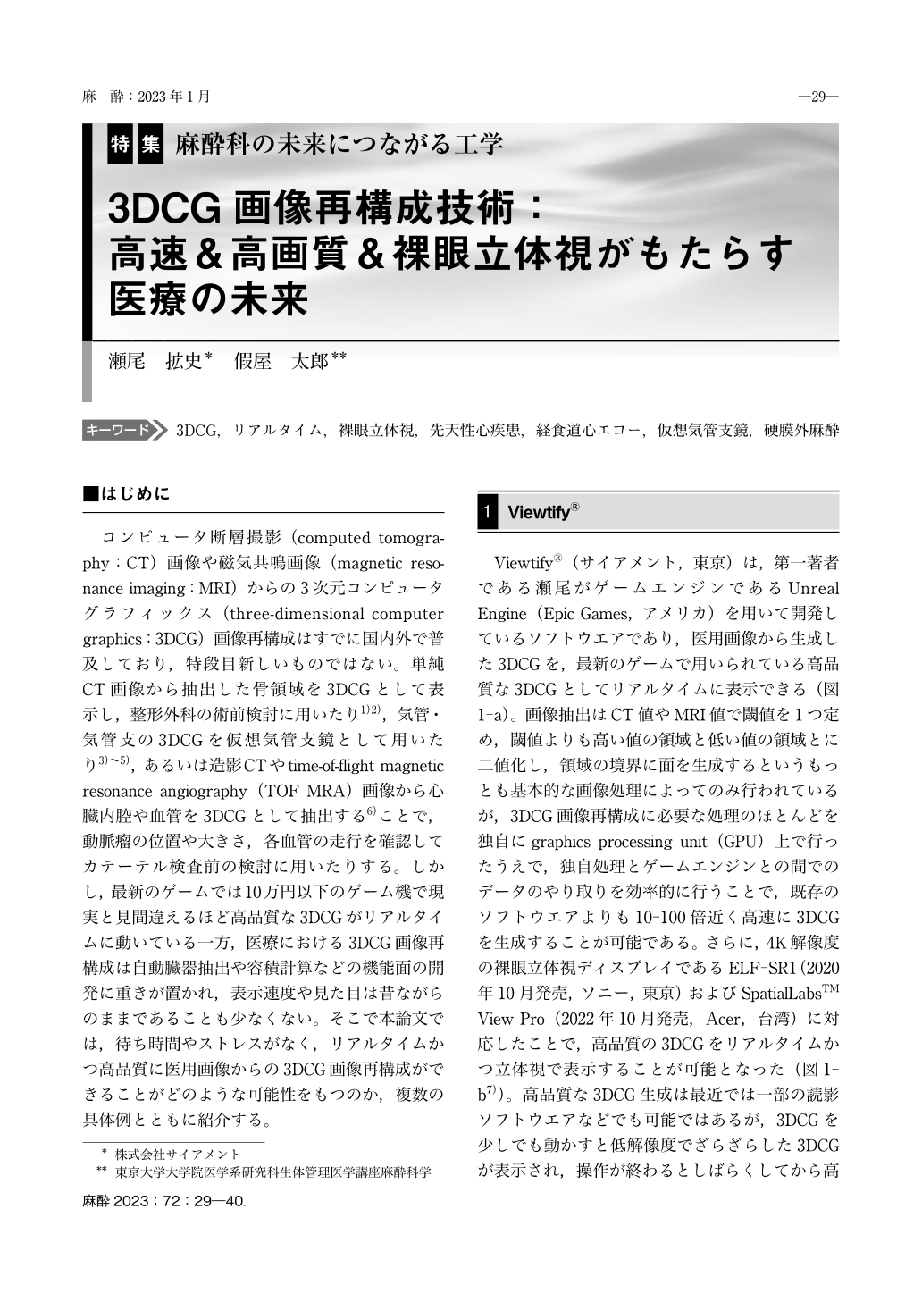
コンピュータ断層撮影(computed tomography:CT)画像や磁気共鳴画像(magnetic resonance imaging:MRI)からの3次元コンピュータグラフィックス(three-dimensional computer graphics:3DCG)画像再構成はすでに国内外で普及しており,特段目新しいものではない。単純CT画像から抽出した骨領域を3DCGとして表示し,整形外科の術前検討に用いたり1)2),気管・気管支の3DCGを仮想気管支鏡として用いたり3)~5),あるいは造影CTやtime-of-flight magnetic resonance angiography(TOF MRA)画像から心臓内腔や血管を3DCGとして抽出する6)ことで,動脈瘤の位置や大きさ,各血管の走行を確認してカテーテル検査前の検討に用いたりする。しかし,最新のゲームでは10万円以下のゲーム機で現実と見間違えるほど高品質な3DCGがリアルタイムに動いている一方,医療における3DCG画像再構成は自動臓器抽出や容積計算などの機能面の開発に重きが置かれ,表示速度や見た目は昔ながらのままであることも少なくない。そこで本論文では,待ち時間やストレスがなく,リアルタイムかつ高品質に医用画像からの3DCG画像再構成ができることがどのような可能性をもつのか,複数の具体例とともに紹介する。
Three-dimensional computer graphics(3DCG)reconstruction from medical images is already common in the medical field, but the appearance and real-time quality of the medical 3DCG have been mostly far from the ones used in the entertainment field, such as computer games. 3DCG reconstruction in the medical field often focuses on the development of algorithms such as automatic organ segmentation with artificial intelligence(AI)or volume calculation, while the speed of 3DCG calculation and appearance often remain the same as in the past. Here we introduce the possibility of real-time, high-quality, naked-eye-stereoscopic 3DCG reconstruction from medical images without waiting time and stress, along with several case examples:the classification of ventricular septal defects, preoperative simulations, transesophageal echocardiography, or virtual bronchoscopy.

Copyright © 2023 KOKUSEIDO CO., LTD. All Rights Reserved.


