Japanese
English
- 有料閲覧
PPV購入案内
1,650円 (1,500円+税10%) こちらは、145ページから162ページの文献です。購入には医書.jp本会員登録が必要です。
会員登録完了後に改めて上記「購入サイトへ」を選択してください。
または「購入サイトへ」選択後に「購入ログイン」、「新規会員登録」の順に進んでください。
会員登録について詳しくはをご確認ください。
- Abstract 文献概要
- 1ページ目 Look Inside
- 参考文献 Reference
- サイト内被引用 Cited by
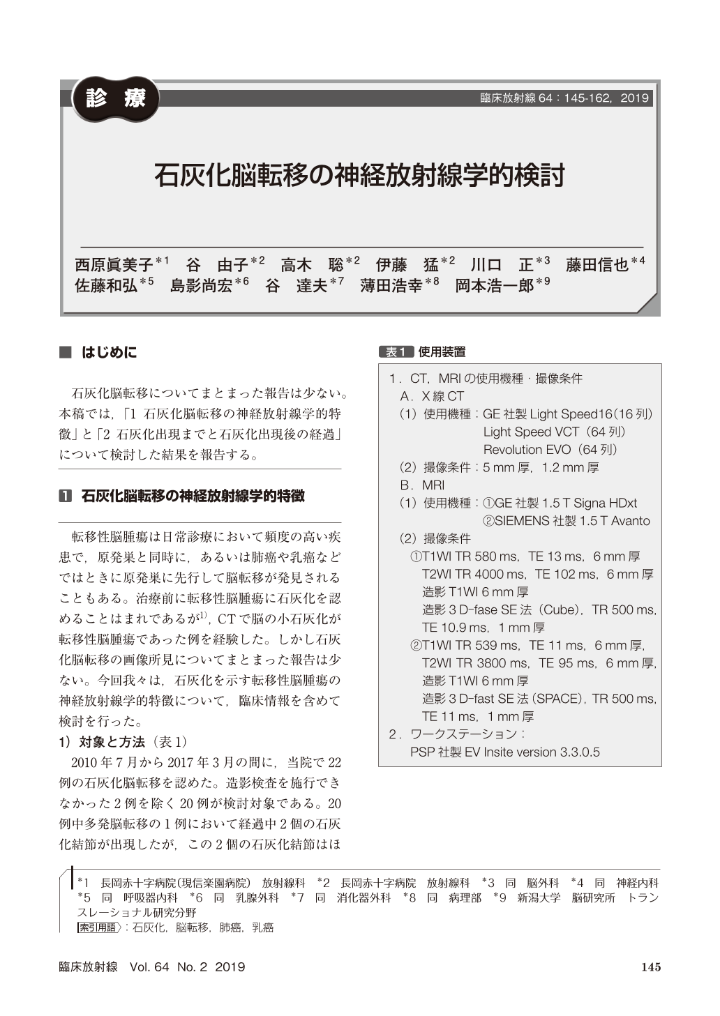
石灰化脳転移についてまとまった報告は少ない。本稿では,「1 石灰化脳転移の神経放射線学的特徴」と「2 石灰化出現までと石灰化出現後の経過」について検討した結果を報告する。
We evaluated clinical and imaging characteristics of 66 calcified brain metastases in the 20 patient(age 31-78 years, median 59 years;male 6 and female 14;adenocarcinoma of the lung 13, large cell neuroendocrine cancer of the lung 1, scirrhous carcinoma of the breast 5, adenocarcinoma of the rectum 1).Contrast effect of the five calcified metastases was unremarkable in three patients initially, although repeated imaging studies revealed significant contrast enhancement. Calcified metastatic brain tumors and the calcification showed variable changes in size and appearance in the clinical course. The calcification persisted after the metastatic tumors disappeared with treatments. Although most calcification developed in the regressed or stable metastatic tumors, calcification could develop in growing metastases.
Cancer metastasis should be considered as a differential diagnosis when small-sized calcifications are seen superficially in the cerebral hemisphere or cerebellum on CT in middle-aged and elderly patients. We recommend MRI study with Gd enhancement and/or screening for malignant tumors, including the lung cancer and breast cancer.
Copyright © 2019, KANEHARA SHUPPAN Co.LTD. All rights reserved.