Japanese
English
- 有料閲覧
- Abstract 文献概要
- 1ページ目 Look Inside
- 参考文献 Reference
Point
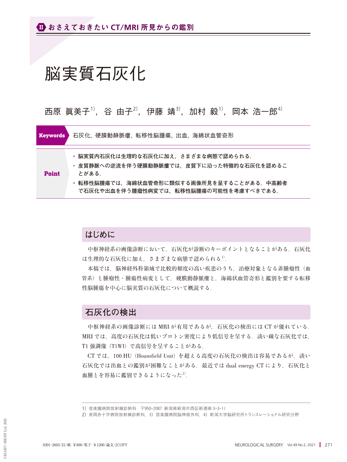
・脳実質内石灰化は生理的な石灰化に加え,さまざまな病態で認められる.
・皮質静脈への逆流を伴う硬膜動静脈瘻では,皮質下に沿った特徴的な石灰化を認めることがある.
・転移性脳腫瘍では,海綿状血管奇形に類似する画像所見を呈することがある.中高齢者で石灰化や出血を伴う腫瘤性病変では,転移性脳腫瘍の可能性を考慮すべきである.
Brain calcification can be either physiological or pathological. Pathological calcification occurs due to a wide spectrum of causes, including congenital disorders, infections, endocrine/metabolic diseases, cerebrovascular diseases, and neoplasms. The patient's age, localization of the calcification, and association with other imaging findings are useful for the correct diagnosis. Dural arteriovenous fistulas with cortical venous reflux should be included in the differential diagnosis of subcortical calcification via CT. MRA should be conducted subsequently. We recently reported the clinical and imaging characteristics of calcified brain metastases in 20 patients. Hemorrhage, necrosis, or degeneration were detected within the lesions in six patients. Both T1WI and T2WI showed a hyperintense mass surrounded by a hypointense rim in one patient. Hemorrhagic brain metastases can mimic cerebral cavernous malformations. Cancer metastasis should be considered as a differential diagnosis when calcified or hemorrhagic masses are detected in middle-aged and elderly patients. We recommend conducting MRI with Gd enhancement.

Copyright © 2021, Igaku-Shoin Ltd. All rights reserved.


