Japanese
English
- 有料閲覧
PPV購入案内
1,320円 (1,200円+税10%) こちらは、445ページから449ページの文献です。購入には医書.jp本会員登録が必要です。
会員登録完了後に改めて上記「購入サイトへ」を選択してください。
または「購入サイトへ」選択後に「購入ログイン」、「新規会員登録」の順に進んでください。
会員登録について詳しくはをご確認ください。
- Abstract 文献概要
- 1ページ目 Look Inside
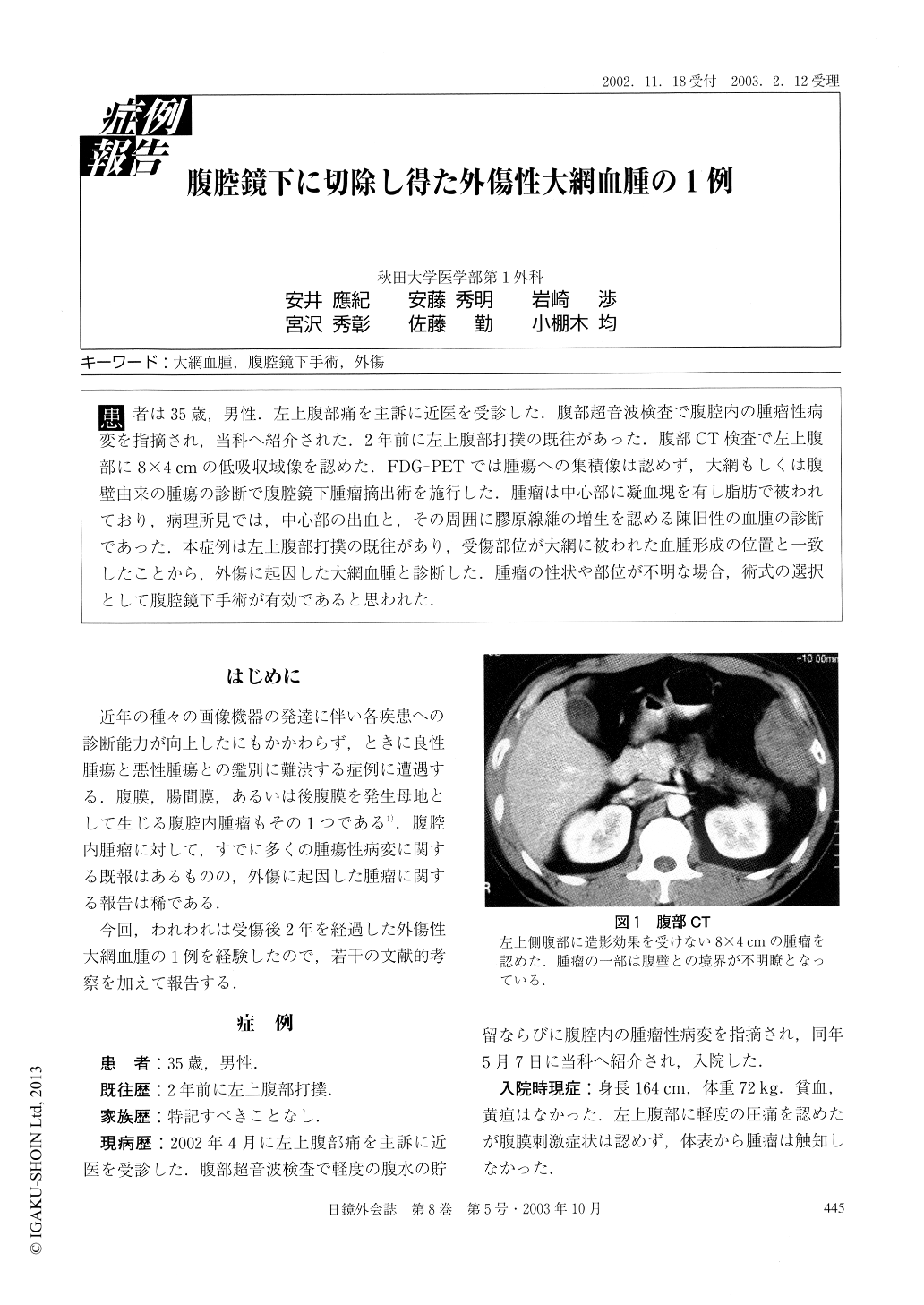
患者は35歳,男性.左上腹部痛を主訴に近医を受診した.腹部超音波検査で腹腔内の腫瘤性病変を指摘され,当科へ紹介された.2年前に左上腹部打撲の既往があった.腹部CT検査で左上腹部に8×4cmの低吸収域像を認めた.FDG-PETでは腫瘍への集積像は認めず,大網もしくは腹壁由来の腫瘍の診断で腹腔鏡下腫瘤摘出術を施行した.腫瘤は中心部に凝血塊を有し脂肪で被われており,病理所見では,中心部の出血と,その周囲に膠原線維の増生を認める陳旧性の血腫の診断であった.本症例は左上腹部打撲の既往があり,受傷部位が大網に被われた血腫形成の位置と一致したことから,外傷に起因した大網血腫と診断した.腫瘤の性状や部位が不明な場合,術式の選択として腹腔鏡下手術が有効であると思われた.
A rare case of a 35-year-old male with trauma-induced omental hematoma is reported. The patient com-plained of abdominal pain and was found to have an intraabdominal mass in the left upper abdomen and ascites. Two years ago he was hospitalized for 2 weeks with contusions on the left upper abdomen. Abdominal CT re-vealed a low density area at the left abdominal wall. PET did not show FDG uptake in the mass. Laparoscopic examination revealed the a mass enveloped in the greater omentum adhered to the left abdominal wall.
Copyright © 2003, JAPAN SOCIETY FOR ENDOSCOPIC SURGERY All rights reserved.