Japanese
English
- 有料閲覧
PPV購入案内
1,320円 (1,200円+税10%) こちらは、993ページから1001ページの文献です。購入には医書.jp本会員登録が必要です。
会員登録完了後に改めて上記「購入サイトへ」を選択してください。
または「購入サイトへ」選択後に「購入ログイン」、「新規会員登録」の順に進んでください。
会員登録について詳しくはをご確認ください。
- Abstract 文献概要
- 1ページ目 Look Inside
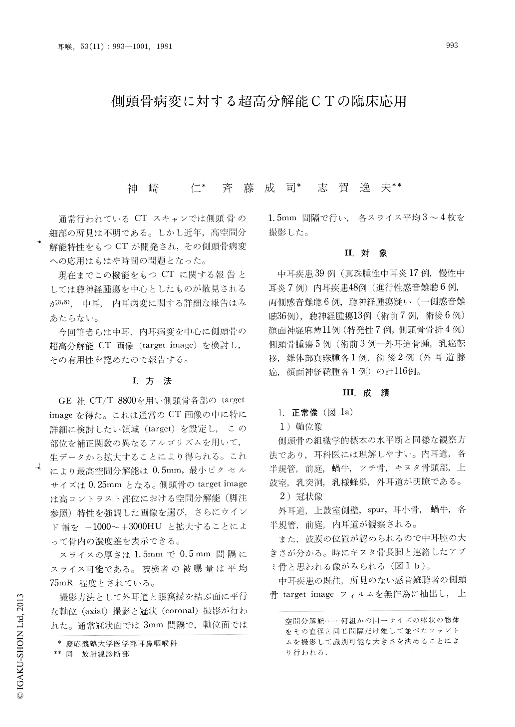
通常行われているCTスキャンでは側頭骨の細部の所見は不明である。しかし近年,高空間分解能特性をもつCTが開発され,その側頭骨病変への応用はもはや時間の問題となった。
現在までこの機能をもつCTに関する報告としては聴神経腫瘍を中心としたものが散見されるが3,8),中耳,内耳病変に関する詳細な報告はみあたらない。
Using CT/T 8800 with high spatial resolution, target imaging of the various temporal bone diseases was performed. Coronal and axial scans were made. Target imaging is an effective means of studying patients with cholesteatomas, semicircular canal fistula, diseases of the internal auditory canal (acoustic neuroma, or stenosis) and inner ear anomaly (hypoplasia, or distension of the vestibulum).
The results of the comparison of CT and polytomography of the temporal bone were as follows: 1. In most cases, the pathological process was demonstrated with CT scans better than or same as with polytomograms. 2. The radiation dose of CT scan is low. 3. The axial images of the temporal bone were easily obtained with CT. 4. CT is more effective than polytomography in the diagnosis and evaluation of the temporal bone involved by tumor and cholesteatoma, especially attic cholesteatoma. 5. In cases with temporal bone fracture, acoustic neuroma or other intracranial diseases, contrast-enhanced CT can be done.
Copyright © 1981, Igaku-Shoin Ltd. All rights reserved.