Japanese
English
- 有料閲覧
PPV購入案内
1,320円 (1,200円+税10%) こちらは、363ページから366ページの文献です。購入には医書.jp本会員登録が必要です。
会員登録完了後に改めて上記「購入サイトへ」を選択してください。
または「購入サイトへ」選択後に「購入ログイン」、「新規会員登録」の順に進んでください。
会員登録について詳しくはをご確認ください。
- Abstract 文献概要
- 1ページ目 Look Inside
I.はじめに
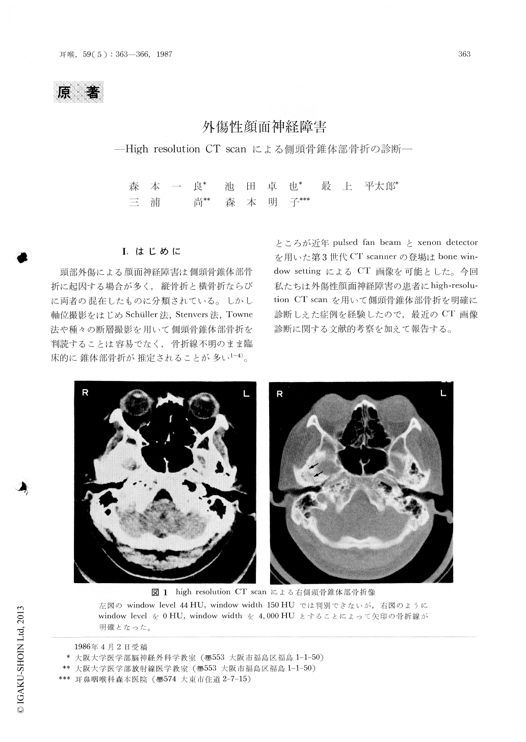
頭部外傷による顔面神経障害は側頭骨錐体部骨折に起因する場合が多く,縦骨折と横骨折ならびに両者の混在したものに分類されている。しかし軸位撮影をはじめSchüller法,Stenvers法,Towne法や種々の断層撮影を用いて側頭骨錐体部骨折を判読することは容易でなく,骨折線不明のまま臨床的に錐体部骨折が推定されることが多い1〜4)。ところが近年pulsed fan beamとxenon detectorを用いた第31世代CT scannerの登場はbone window settingによるCT画像を可能とした。今回私たちは外傷性顔面神経障害の患者にhigh-resolution CT scanを用いて側頭骨錐体部骨折を明確に診断しえた症例を経験したので,最近のCT画像診断に関する文献的考察を加えて報告する。
Facial paralysis can be caused by severe head injury and trauma of temporal region of the skull. A case of facial paralysis with absence of lacrimation due to transverse fracture of the petrous bone was reported.
We confirmed the transverse fracture of the pyramis which was suspected with conventional radiological study by high-resolution, thin section CT scan. High-resolution, thin section CT scan is effective in detecting the bony detail and fracture line that passes through the petrous bone in terms of great accuracy and sensitivity.
Copyright © 1987, Igaku-Shoin Ltd. All rights reserved.