Japanese
English
- 有料閲覧
- Abstract 文献概要
- 1ページ目 Look Inside
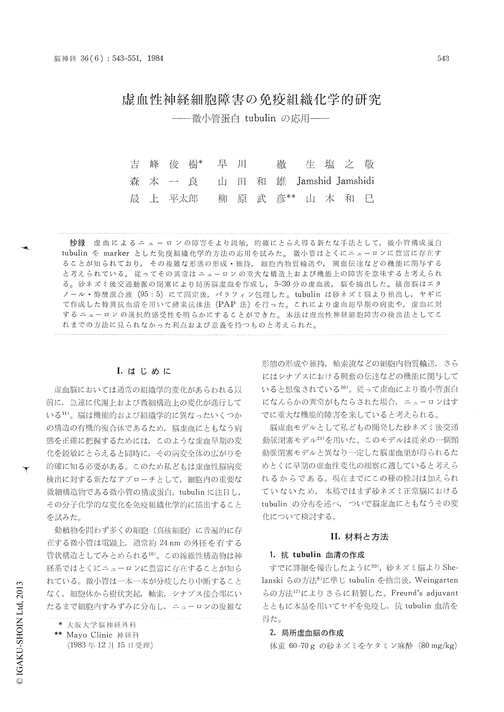
抄録 虚血によるニューロンの障害をより鋭敏,的確にとらえ得る新たな手法として,微小管構成蛋白tubulinをmarkerとした免疫組織化学的方法の応用を試みた。微小管はとくにニューロンに豊富に存在することが知られており,その複雑な形態の形成・維持,細胞内物質輸送や,興奮伝達などの機能に関与すると考えられている。従ってその異常はニューロンの重大な構造上および機能上の障害を意味すると考えられる。砂ネズミ後交通動脈の閉塞により局所脳虚血を作成し,5-30分の虚血後,脳を摘出した。摘出脳はエタノール。酢酸混合液(95:5)にて固定後,パラフィン包埋した。tubulinは砂ネズミ脳より抽出し,ヤギにて作成した特異抗血清を用いて酵素抗体法(PAP法)を行った。これにより虚血超早期の病変や,虚血に対するニューロンの選択的感受性を明らかにすることができた。本法は虚血性神経細胞障害の検出法としてこれまでの方法に見られなかった利点および意義を持つものと考えられた。
The limitation of conventional histological me-thods to demonstrate ischemic change of neurons in early phase has been a major drawback in histopathological and pathophysiological studies of cerebral ischemia. Cellular metabolism is disturb-ed immediately after cessation of the regional circulation and rapid alterations in macromolecular and ultrastructural integrity in neurons may take place before any evidence of histopathological changes could be detectable. To demonstrate ischemic change of neurons more sensitively on a histological level, we applied immunohistochem-ical method using antiserum to tubulin, a protein of microtubules. As this organelle has been implicated in several important cellular functions such as control of cell shape, intracytoplasmic transport of materials or synaptic transduction, immunohistochemical alterations in microtubules may indicate structural as well as functional damage of neurons.
In order to study the ischemic change in neu-rons, the posterior communicating artery of a gerbil brain was occluded by the method pre-viously reported by us, and the hippocampus, which is one of the most vulnerable structures of the brain to ischemia, was observed. Five or 30 minutes after occlusion, animals were sacrificed by decapitation. Brains were removed, cut coron-ally and fixed in ethanol-acetic acid (95:5). Tubulin used for this study was extracted from normal gerbil brains and specific antiserum was raised in goats Peroxidase-antiperoxidase method was performed on paraffin sections. Immunohisto-chemical distribution of tubulin in a normal gerbil brain demonstrated by the present method was in good accordance with the reported electron-microscopical distribution of microtubules. Five minutes after ischemia, a small number of pyra-midal cells in the CA 1 area of the hippocampuslost their reaction to the antiserum on the oc-cluded side whereas neurons on the contralateral hippocampus remained normal. At 30 minutes of ischemia, most neurons in the CA 1 area lost their reaction to the antiserum. However, a few neurons in stratum oriens and the pyramidal cell layer remained positive for tubulin staining. Dif-ference in vulnerability to ischemia between these interneurons and pyramidal cells was discussed. The mechanism of loss of tubulin stainability in ischemic neurons was discussed on chemical and immunohistochmical basis. At present, we spe-culate that the disturbance in calcium homeostasis in ischemic cells could be an essential factor to depolymerize microtubular tubulin and to reduce immunohistochemical reactivity.
The present investigation demostrated that the immunohistochemical method with antiserum to tubulin is a useful tool to observe chemical or molecular change of the microtubular protein on a histological level and to study neuronal damage to ischemia.

Copyright © 1984, Igaku-Shoin Ltd. All rights reserved.


