Japanese
English
- 有料閲覧
- Abstract 文献概要
- 1ページ目 Look Inside
I.はじめに
MRIの検査時間が短縮されたとは言え,体内金属の有無を含め,詳しく患者の状況を把握しえていない初診においては5),急な意識障害,片麻痺,失語などで発症した頭蓋内疾患が疑われる症例に対しては,まずCTが施行されることが一般的である.
発症後間もない脳梗塞は,初診時のCTではその確定診断となる低吸収域を認めることは少なく,半日あるいは一日あいだをあけてのCTやMRIの再検にて確定診断に至ることも実際の診療現場においては少なくない.
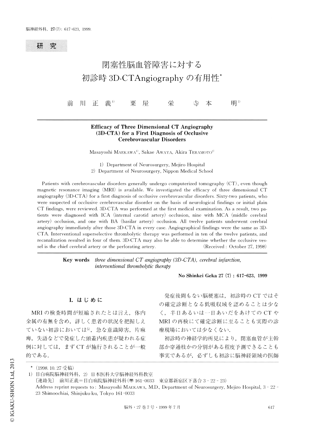
Patients with cerebrovascular disorders generally undergo computerized tomography (CT), even thoughmagnetic resonance imaging (MRI) is available. We investigated the efficacy of three dimensional CTangiography (3D-CTA) for a first diagnosis of occlusive cerebrovascular disorders. Sixty-two patients, whowere suspected of occlusive cerebrovascular disorder on the basis of neurological findings or initial plainCT findings, were reviewed. 3D-CTA was performed at the first medical examination. As a result, two pa-tients were diagnosed with ICA (internal carotid artery) occlusion, nine with MCA (middle cerebralartery) occlusion, and one with BA (basilar artery) occlusion. All twelve patients underwent cerebralangiography immediately after those 3D-CTA in every case. Angiographical findings were the same as 3D-CTA. Interventional superselective thrombolytic therapy was performed in ten of the twelve patients, andrecanalization resulted in four of them. 3D-CTA may also be able to determine whether the occlusive ves-sel is the chief cerebral artery or the perforating artery.

Copyright © 1999, Igaku-Shoin Ltd. All rights reserved.


