Japanese
English
- 有料閲覧
PPV購入案内
1,320円 (1,200円+税10%) こちらは、237ページから243ページの文献です。購入には医書.jp本会員登録が必要です。
会員登録完了後に改めて上記「購入サイトへ」を選択してください。
または「購入サイトへ」選択後に「購入ログイン」、「新規会員登録」の順に進んでください。
会員登録について詳しくはをご確認ください。
- Abstract 文献概要
- 1ページ目 Look Inside
I.はじめに
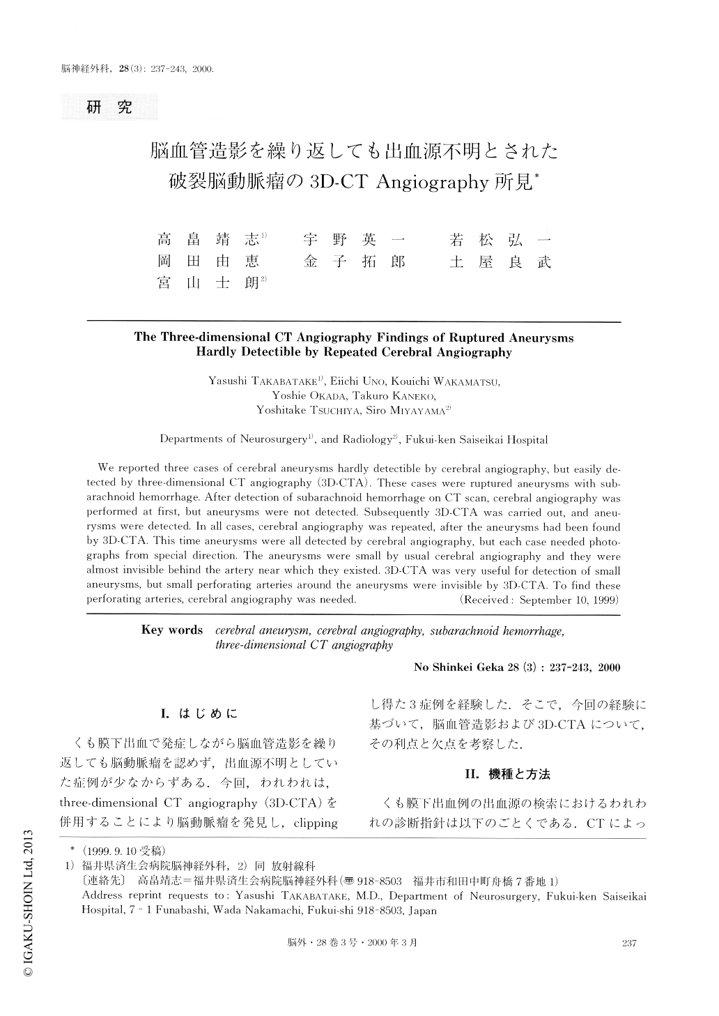
くも膜下出血で発症しながら脳血管造影を繰り返しても脳動脈瘤を認めず,出血源不明としていた症例が少なからずある.今回,われわれは,three-dimensional CT angiography(3D-CTA)を併用することにより脳動脈瘤を発見し,clippingし得た3症例を経験した.そこで,今回の経験に基づいて,脳血管造影および3D-CTAについて,その利点と欠点を考察した.
We reported three cases of cerebral aneurysms hardly detectible by cerebral angiography, but easily de-tected by three-dimensional CT angiography (3D-CTA). These cases were ruptured aneurysms with sub-arachnoid hemorrhage. After detection of subarachnoid hemorrhage on CT scan, cerebral angiography was performed at first, but aneurysms were not detected. Subsequently 3D-CTA was carried out, and aneu-rysms were detected. In all cases, cerebral angiography was repeated, after the aneurysms had been found by 3D-CTA. This time aneurysms were all detected by cerebral angiography, but each case needed photo-graphs from special direction. The aneurysms were small by usual cerebral angiography and they were almost invisible behind the artery near which they existed. 3D-CTA was very useful for detection of small aneurysms, but small perforating arteries around the aneurysms were invisible by 3D-CTA. To find these perforating arteries, cerebral angiography was needed.
Copyright © 2000, Igaku-Shoin Ltd. All rights reserved.