Japanese
English
- 有料閲覧
- Abstract 文献概要
- 1ページ目 Look Inside
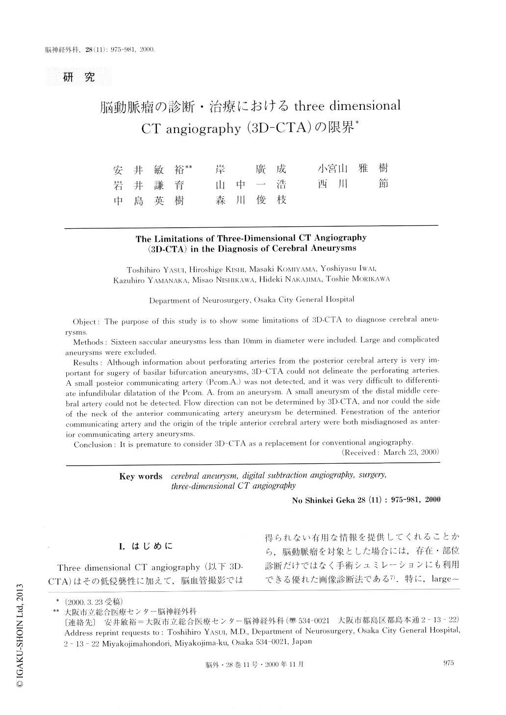
I.はじめに
Three dimensional CT angiography(以下3D-CTA)はその低侵襲性に加えて,脳血管撮影では得られない有用な情報を提供してくれることから,脳動脈瘤を対象とした場合には,存在・部位診断だけではなく手術シュミレーションにも利用できる優れた画像診断法である7).特に,large〜giant7)あるいは紡錘形15)などの特殊な動脈瘤の治療戦略を考える場合には,必須の診断法と考えられる.筆者らも1993年の開院以来,未破裂脳動脈瘤症例を中心として,脳血管撮影(当院ではDi-gital subtraction angiographyのため,以下DSA)と併用して3D-CTAを行ってきた.その後,徐々に3D-CTAによる脳動脈瘤の診断に習熟するにつれて,通常サイズの嚢状動脈瘤の場合には術前検査として,DSAは行わずMR angio-graphy(以下,MRA)と3D-CTAのみで手術を行うようになってきた.しかし,3D-CTA,MRAのみでは十分な情報が得られず,DSAの必要性を感じる症例も経験するようになってきた.今回はこれまでに経験した問題例を提示し,脳動脈瘤の診断における3D-CTAの限界について報告する.
Object: The purpose of this study is to show some limitations of 3D-CTA to diagnose cerebral aneu-rysms.
Methods: Sixteen saccular aneurysms less than 10mm in diameter were included. Large and complicated aneurysms were excluded.
Results: Although information about perforating arteries from the posterior cerebral artery is very im-portant for sugery of basilar bifurcation aneurysms, 3D-CTA could not delineate the perforating arteries. A small posteior communicating artery (Pcom.A.) was not detected, and it was very difficult to differenti-ate infundibular dilatation of the Pcom. A. from an aneurysm. A small aneurysm of the distal middle cere-bral artery could not be detected. Flow direction can not be determined by 3D-CTA, and nor could the side of the neck of the anterior communicating artery aneurysm be determined. Fenestration of the anterior communicating artery and the origin of the triple anterior cerebral artery were both misdiagnosed as anter-ior communicating artery aneurysms.
Conclusion: It is premature to consider 3D-CTA as a replacement for conventional angiography.

Copyright © 2000, Igaku-Shoin Ltd. All rights reserved.


