Japanese
English
- 有料閲覧
- Abstract 文献概要
- 1ページ目 Look Inside
- 参考文献 Reference
Ⅰ.はじめに
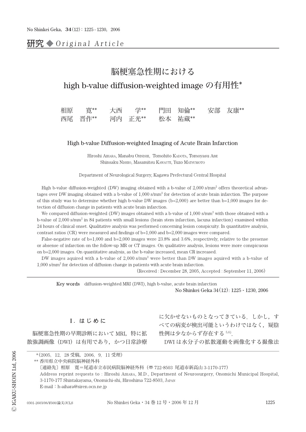
脳梗塞急性期の早期診断においてMRI,特に拡散強調画像(DWI)は有用であり,かつ日常診療に欠かせないものとなってきている.しかし,すべての病変が検出可能というわけではなく,疑陰性例は少なからず存在する5,6).
DWIは水分子の拡散運動を画像化する撮像法で,その拡散強調の程度はb値によって表現される.一般的には,b値は1,000s/mm2程度が用いられている.最近の強力な傾斜磁場を発生できるMRI装置は,b値をより高くし,より高度な拡散強調を行った撮像(high b-value DWI)を容易に施行可能とし,脳梗塞急性期における病変と正常組織との間のコントラストが高くなり,病変検出能は上がると考えられる.
当院ではDWI施行可能なMRI装置を2002年4月に導入し,脳梗塞急性期のMRIとして当初はT2WI,FLAIR,TOF-MRA,b=1,000s/mm2のDWIを施行していたが,偽陰性例が散見されるようになり,b=2,000s/mm2のDWIを追加するようになった.
今回われわれは,脳梗塞急性期にb=1,000s/mm2で撮像した通常のDWI(DWIb=1000)よりもb=2,000s/mm2で撮像したhigh b-value DWI(DWIb=2000)が病変検出能を上げることができるかどうかを調査した.
High b-value diffusion-weighted (DW) imaging obtained with a b-value of 2,000s/mm2 offers theoretical advantages over DW imaging obtained with a b-value of 1,000s/mm2 for detection of acute brain infarction. The purpose of this study was to determine whether high b-value DW images (b=2,000) are better than b=1,000 images for detection of diffusion change in patients with acute brain infarction.
We compared diffusion-weighted (DW) images obtained with a b-value of 1,000 s/mm2 with those obtained with a b-value of 2,000s/mm2 in 84 patients with small lesions (brain stem infarction,lacuna infarction) examined within 24 hours of clinical onset. Qualitative analysis was performed concerning lesion conspicuity. In quantitative analysis,contrast ratios (CR) were measured and findings of b=1,000 and b=2,000 images were compared.
False-negative rate of b=1,000 and b=2,000 images were 23.8% and 3.6%,respectively,relative to the presense or absense of infarction on the follow-up MR or CT images. On qualitative analysis,lesions were more conspicuous on b=2,000 images. On quantitative analysis,as the b-value increased,mean CR increased.
DW images aquired with a b-value of 2,000s/mm2 were better than DW images aquired with a b-value of 1,000s/mm2 for detection of diffusion change in patients with acute brain infarction.

Copyright © 2006, Igaku-Shoin Ltd. All rights reserved.


