Japanese
English
- 有料閲覧
PPV購入案内
1,320円 (1,200円+税10%) こちらは、359ページから365ページの文献です。購入には医書.jp本会員登録が必要です。
会員登録完了後に改めて上記「購入サイトへ」を選択してください。
または「購入サイトへ」選択後に「購入ログイン」、「新規会員登録」の順に進んでください。
会員登録について詳しくはをご確認ください。
- Abstract 文献概要
- 1ページ目 Look Inside
I.緒言
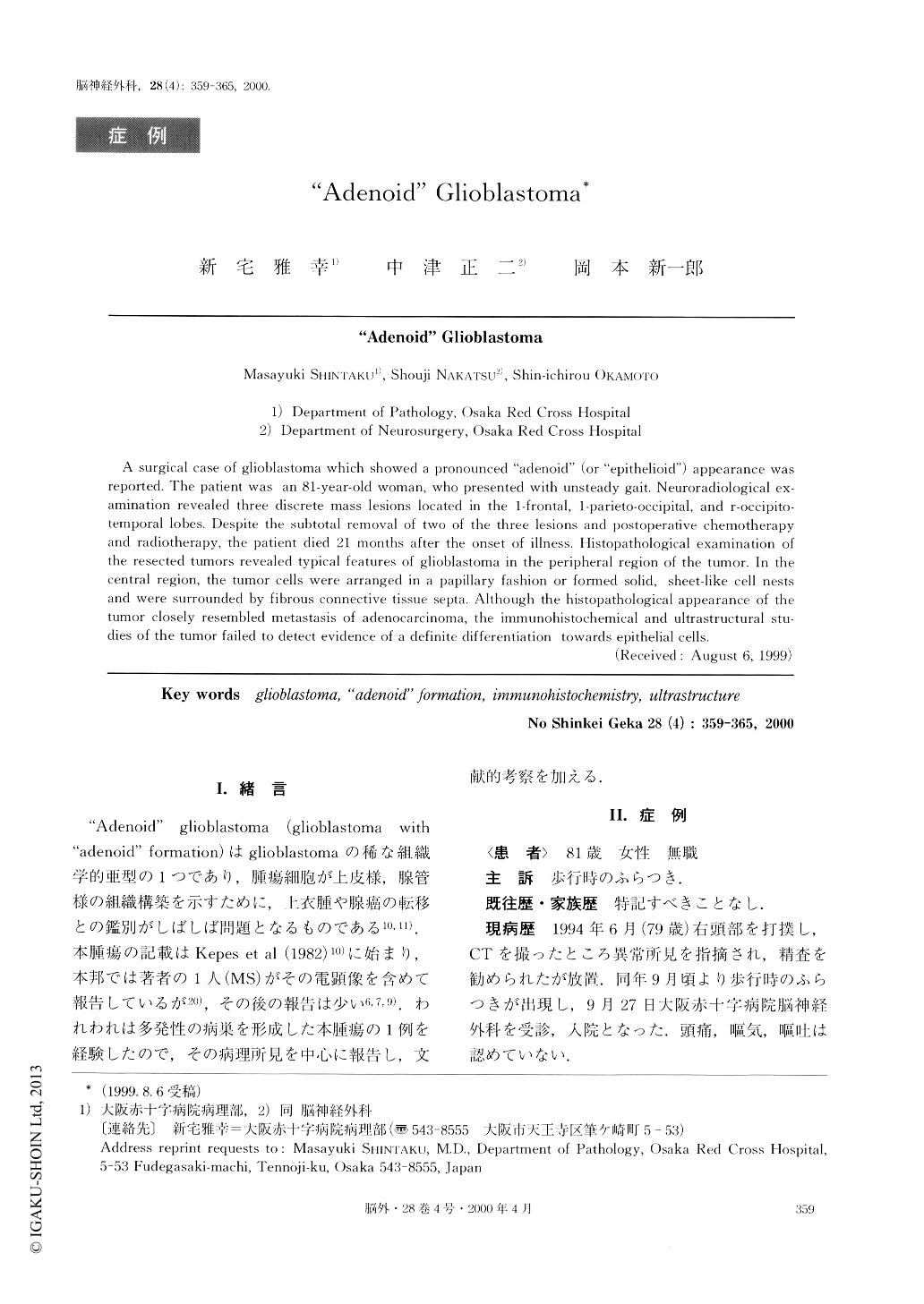
“Adenoid”glioblastoma(glioblastoma with“adenoid”formation)はglioblastomaの稀な組織学的亜型の1つであり,腫瘍細胞が上皮様,腺管様の組織構築を示すために,上衣腫や腺癌の転移との鑑別がしばしば問題となるものである10,11).本腫瘍の記載はKepes et al(1982)10)に始まり,本邦では著者の1人(MS)がその電顕像を含めて報告しているが20),その後の報告は少い6,7,9).われわれは多発性の病巣を形成した本腫瘍の1例を経験したので,その病理所見を中心に報告し,文献的考察を加える.
A surgical case of glioblastoma which showed a pronounced “adenoid” (or “epithelioid”) appearance was reported. The patient was an 81-year-old woman, who presented with unsteady gait. Neuroradiological ex-amination revealed three discrete mass lesions located in the 1-frontal, 1-parieto-occipital, and r-occipito-temporal lobes. Despite the subtotal removal of two of the three lesions and postoperative chemotherapy and radiotherapy, the patient died 21 months after the onset of illness. Histopathological examination of the resected tumors revealed typical features of glioblastoma in the peripheral region of the tumor. In the central region, the tumor cells were arranged in a papillary fashion or formed solid, sheet-like cell nests and were surrounded by fibrous connective tissue septa. Although the histopathological appearance of the tumor closely resembled metastasis of adenocarcinoma, the immunohistochemical and ultrastructural stu-dies of the tumor failed to detect evidence of a definite differentiation towards epithelial cells.
Copyright © 2000, Igaku-Shoin Ltd. All rights reserved.