Japanese
English
- 有料閲覧
PPV購入案内
1,320円 (1,200円+税10%) こちらは、889ページから894ページの文献です。購入には医書.jp本会員登録が必要です。
会員登録完了後に改めて上記「購入サイトへ」を選択してください。
または「購入サイトへ」選択後に「購入ログイン」、「新規会員登録」の順に進んでください。
会員登録について詳しくはをご確認ください。
- Abstract 文献概要
- 1ページ目 Look Inside
I.はじめに
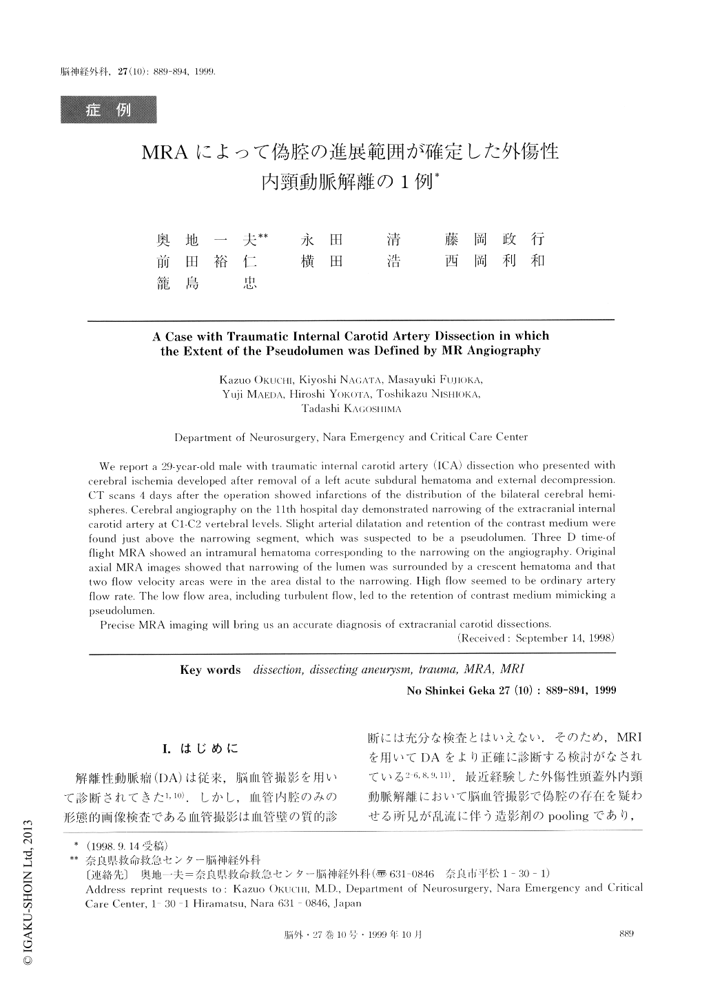
解離性動脈瘤(DA)は従来,脳血管撮影を用いて診断されてきた1,10).しかし,血管内腔のみの形態的画像検査である血管撮影は血管壁の質的診断には充分な検査とはいえない.そのため,MRIを用いてDAをより正確に診断する検討がなされている2-6,8,9,11).最近経験した外傷性頭蓋外内頸動脈解離において脳血管撮影で偽腔の存在を疑わせる所見が乱流に伴う造影剤のpoolingであり,MRAを用いて正確な解離の進展範囲を診断できた1例を経験したので報告する.
We report a 29-year-old male with traumatic internal carotid artery (ICA) dissection who presented withcerebral ischemia developed after removal of a left acute subdural hematoma and external decompression.CT scans 4 days after the operation showed infarctions of the distribution of the bilateral cerebral hemi-spheres. Cerebral angiography on the 11th hospital day demonstrated narrowing of the extracranial internalcarotid artery at C1-C2 vertebral levels. Slight arterial dilatation and retention of the contrast medium werefound just above the narrowing segment, which was suspected to be a pseudolumen. Three D time-of flight MRA showed an intramural hematoma corresponding to the narrowing on the angiography. Originalaxial MRA images showed that narrowing of the lumen was surrounded by a crescent hematoma and thattwo flow velocity areas were in the area distal to the narrowing. High flow seemed to be ordinary artery flow rate. The low flow area, including turbulent flow, led to the retention of contrast medium mimicking apseudolumen.
Precise MRA imaging will bring us an accurate diagnosis of extracranial carotid dissections.
Copyright © 1999, Igaku-Shoin Ltd. All rights reserved.