Japanese
English
- 有料閲覧
- Abstract 文献概要
- 1ページ目 Look Inside
I.はじめに
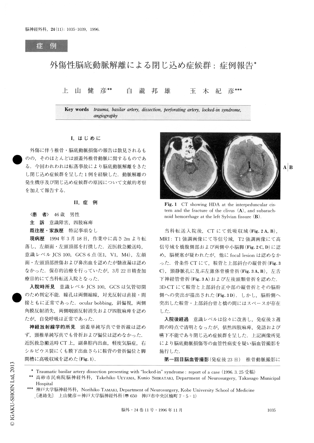
外傷に伴う椎骨・脳底動脈損傷の報告は散見されるものの,そのほとんどは頭蓋外椎骨動脈に関するものである.今回われわれは転落事故により脳底動脈解離をきたし閉じ込め症候群を呈した1例を経験した.動脈解離の発生機序及び閉じ込め症候群の原因について文献的考察を加えて報告する.
We encountered a case of traumatic basilar artery dissection presenting with “locked-in” syndrome.
A 46-year-old-man fell approximately 2 meters and landed in an inverted position. He was admitted to our hospital after 4 days. He was in coma on admission, and neurological examination revealed that his pupils were 2mm in size and reactive, the corneal reflex of both sides was absent, the pharyngeal reflex was ab-sent, and quadriplegia was present. CT and MRI showed infarction at the ventral portion of the pons.3D-CT and CT using a bone algorithm showed frac-tures of the upper clivus, the left petrous bone, and the left occipital condyle. Angiography showed a pearl and string sign at the upper basilar artery. It is suggested that the cause of the infarction of the ventral portion of the pons was the occlusion of the perforating arteries due to the basilar artery dissection, and the mechanism of the basilar artery dissection was the hyperextension or heperflexion of the basilar artery by the movement of the brain stem caused by the traumatic impact. Re-peated angiography revealed gradual improvement. Eventually he freed himself from the “locked-in” syn-drome.

Copyright © 1996, Igaku-Shoin Ltd. All rights reserved.


